Tension Headaches
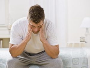
New research shows that spinal manipulation may be an effective treatment option for tension headaches and headaches that originate in the neck.
What You Should Know
If you have a headache, you’re not alone. Nine out of ten Americans suffer from headaches. Some are occasional, some frequent, some are dull and throbbing, and some cause debilitating pain and nausea.
+ Read More
What do you do when you suffer from a pounding headache? Do you grit your teeth and carry on? Lie down? Pop a pill and hope the pain goes away? There is a better alternative. New research shows that spinal manipulation – the primary form of care provided by doctors of chiropractic – may be an effective treatment option for tension headaches and headaches that originate in the neck. A report released in 2001 by researchers at the Duke University Evidence-Based Practice Center in Durham, NC, found that spinal manipulation resulted in almost immediate improvement for those headaches that originate in the neck, and had significantly fewer side effects and longer-lasting relief of tension-type headache than a commonly prescribed medication. Also, a 1995 study in the Journal of Manipulative and Physiological Therapeutics found that spinal manipulative therapy is an effective treatment for tension headaches , and that those who ceased chiropractic treatment after four weeks experienced a sustained therapeutic benefit in contrast with those patients who received a commonly prescribed medication.
Causes
To get to the bottom of the problem, you first need to find out what is causing your pain. Headaches have many causes or “triggers.” These may include foods, environmental stimuli (noises, lights, stress, etc.) and/or behaviors (insomnia, excessive exercise, blood sugar changes, etc.). About 5 percent of all headaches are warning signals caused by physical problems. Ninety-five percent of headaches are primary headaches, such as tension, migraine, or cluster headaches. These types of headaches are not caused by disease. The headache itself is the primary concern. “The greatest majority of primary headaches are associated with muscle tension in the neck,” says Dr. George B. McClelland, a doctor of chiropractic from Christiansburg, VA, and chairman of the American Chiropractic Association’s (ACA) Board of Governors. “Today, Americans engage in more sedentary activities than they used to, and more hours are spent in one fixed position or posture. This can increase joint irritation and muscle tension in the neck, upper back and scalp, causing your head to ache.”
What Can You Do?
The American Chiropractic Association’s (ACA) suggests the following: If you spend a large amount of time in one fixed position, such as in front of a computer, on a sewing machine, typing, or reading, take a break and stretch every 30 minutes to one hour. The stretches should take your head and neck through a comfortable range of motion. Low-impact exercise may help relieve the pain associated with primary headaches. However, if you are prone to dull, throbbing headaches, avoid heavy exercise. Engage in such activities as walking and low-impact aerobics . Avoid teeth clenching. This results in stress at the temporomandibular joints (TMJ) – the two joints that connect your jaw to your skull – leading to TMJ irritation and a form of tension headaches. Drink at least eight 8-ounce glasses of water a day to help avoid dehydration, which can lead to headaches. In addition, the ACA and its Council on Nutrition suggest you avoid the following food “triggers”: Avoid caffeine. Foods such as chocolate, coffee, sodas and cocoa contain high levels of the stimulant. Avoid foods with a high salt or sugar content. These foods may cause migraines, resulting in sensitivity to light, noise, or abrupt movements. Avoid drinking alcoholic beverages. These drinks can dehydrate you and cause headache pain. Other headache sufferers may want to avoid not only caffeine, but also high-protein foods, dairy products, red meat and salty foods.
What Can a Doctor of Chiropractic Do?
Dr. McClelland says your doctor of chiropractic may do one or more of the following if you suffer from a primary headache: Perform spinal manipulation or chiropractic adjustments to improve spinal function and alleviate the stress on your system. Provide nutritional advice, recommending a change in diet and perhaps the addition of B complex vitamins. Offer advice on posture, ergonomics (work postures), exercises and relaxation techniques. This advice should help to relieve the recurring joint irritation and tension in the muscles of the neck and upper back. “Doctors of chiropractic undergo extensive training to help their patients in many ways– not just back pain,” says Dr. McClelland. “They know how tension in the spine relates to problems in other parts of the body, and they can take steps to relieve those problems.” If your headache is symptomatic of a health problem that needs the care of another discipline, your doctor of chiropractic will refer you to an appropriate specialist.
Migraine Headaches

Migraines, which usually begin sometime between the teen years and the age of 40, can be classified as either “classic” or “common.”
What You Should Know
Migraine headaches typically affect one side of the head. They can last anywhere from a few hours to a few days. Some people get them weekly, others have fewer than one a year. Migraines, which usually begin sometime between the teen years and the age of 40, can be classified as either “classic” or “common.” Migraine headaches can often leave the recipient feeling depressed, out of control, and totally overwhelmed.
+ Read More
Causes
Recent scientific developments are providing some answers to the cause. What the researchers are discovering is that migraines are not just a headache. Migraines were once believed to be a disorder of anxious, neurotic women whose blood vessels overreacted. While there is a vascular component, migraines are most likely neurobiological disorders of the brain. There is still much to discover and understand about migraines; however, progress is being made.
Signs/Symptoms
You may know you are going to have a migraine before the headache starts. Warning signs include nausea, vomiting, and sensitivity to noise, light, or smells. Classic migraines begin with warning signs such as flashing lights or colors. You may feel as though you are looking through a tunnel. One side of your body may feel prickly, hot, or weak. These warning signs last about 15 to 30 minutes and are followed by pain in your head. Common migraines do not have the same warning signs. However, you may feel tired, depressed, restless, or talkative for 2 or 3 days before the headache starts.
What Can You Do?
At Holmes Spine & Sport Chiropractic, we are dedicated to providing relief from your migraines. It’s believed 70-80% of migraines are triggered from upper neck problems that affect the lower motor neurons that in turn affect the upper motor neurons in the brain leading to migraines. If we can correct problems in the cervical spine you can decrease or potentially eliminate your migraines.
Our treatment guidelines are as follows
- Corrective chiropractic treatment of the spine and temporal mandibular joints (TMJ) using chiropractic, massage, and exercise rehab.
- Medications. Moderation is the key, many of them can cause rebound headaches if taken too frequently. Be aware of the potential for rebound with each medication you take. (Medications will need to be prescribed by your MD.)
- Avoid triggers. Use a headache diary to identify triggers such as certain foods, stress, postural positions, time of day, month, or year, lack of sleep, etc.
Cervical Degenerative Disc Disease
Cervical disc degeneration is a common cause of neck pain
Disc degeneration
Cervical disc degeneration is a common cause of neck pain, most frequently felt as a stiff neck. Cervical degenerative disc disease is much less common than disc degeneration in the lumbar spine because the neck generally is subjected to far less torque and force. Nonetheless, a fall, whiplash or a twisting injury to the disc space can spur degeneration, and accumulated wear and tear on the disc over time can also lead to neck pain caused by disc degeneration.
+ See More +
Cervical degenerative disc disease pain and symptoms
In addition to having the low-grade pain of a stiff or inflexible neck, many patients with cervical disc degeneration have numbness, tingling, or even weakness in the neck, arms, or shoulders as a result of nerves in the cervical area becoming irritated or pinched. For example, a pinched nerve root in the C6-C7 segment could result in weakness in the triceps and forearms, wrist drop and altered sensation in the middle fingers or fingertips. Cervical disc degeneration can also contribute to spinal stenosis, and other progressive conditions, as well as a more sudden disc herniation.
Cervical degenerative disc disease diagnosis
Successful diagnosis of cervical degenerative disc disease begins with a physician reviewing the patient’s history of symptoms and performing a physical examination to measure neck extension and flexibility. During the exam, patients may be asked to perform certain movements and report whether the neck pain increases or decreases. If a physical exam warrants further investigation, imaging studies such as X-Ray, MRI and possibly a CT scan will be taken. These diagnostic images can confirm whether and where degeneration is occurring, and can identify other conditions (such as calcification or arthritis) that could be causing the symptoms.
Cervical degenerative disc disease treatment
The general treatment is largely the same as for degenerative disc disease in the lumbar spine. That is, conservative care (no-surgical) is recommended as the primary strategy and surgery is only considered if a concerted effort at conservative care fails to provide adequate pain relief or a patient’s daily activity has been significantly compromised.
- Chiropractic manipulation can relieve low back pain by taking pressure off sensitive nerves or tissue, increasing range of motion, restoring blood flow, reducing muscle tension, and, like more active exercise, promoting the release of endorphins within the body to act as natural painkillers
- Over the counter and prescription medications may provide relief. These include non-steroidal anti-inflammatories (NSAIDs) and pain relievers like acetaminophen (such as Tylenol). Prescription medications such as oral steroids, muscle relaxants or narcotic pain medications may also be used.
- Exercise, specifically stretching as many dimensions of the neck as possible,is essential to maintain flexibility in the neck and relieve chronic stiffness. A specific set of exercises should be developed by a physician or physical therapist. Some exercises that could be done several times a day include:
- Chin-to-chest stretch, which stretches the back of the neck
- Side-to-side swivel, which involves slowly turning the head to the left and right
- Eyes-to-the-sky, where a patient lifts the chin upward to stretch the front of the neck and upper thoracic area
- Ear-to-shoulder stretch to extend the sides of the neck as much as possible (this can be facilitated by gently placing a hand on the head but should not involve pulling or pushing the neck and head to the shoulder)
- Use of a cervical pillows or neck traction may also be recommended to stabilize the neck and improve neck alignment so the disc compression is not exacerbated as a patient sleeps or relaxes at home
Surgery
If pain is not relieved adequately or daily activities become difficult, surgery may be considered.
Cervical Herniated Disc
Cervical herniated disc symptoms and treatment options Cervical herniated disc introduction Arm pain from a cervical herniated disc is one of the more common cervical spine conditions treated by chiorpactors. It usually develops in the 30 – 50 year old age group. Although a cervical herniated disc may originate from some sort of trauma or injury to the cervical spine, the symptoms, including arm pain, commonly start spontaneously.
+ See More +
The arm pain from a cervical herniated disc results because the herniated disc material “pinches” or presses on a cervical nerve, causing pain to radiate along the nerve pathway down the arm. Along with the arm pain, numbness and tingling can be present down the arm and into the fingertips. Muscle weakness may also be present due to a cervical herniated disc. The two most common levels in the cervical spine to herniate are the C5 – C6 level (cervical 5 and cervical 6) and the C6 -C7 level. The next most common is the C4 – C5 level, and rarely the C7 – T1 level may herniate. The nerve that is affected by the cervical disc herniation is the one exiting the spine at that level, so at the C5-C6 level it is the C6 nerve root that is affected. Symptoms of a cervical herniated disc A cervical herniated disc will typically cause pain patterns and neurological deficits as follows: C4 – C5 (C5 nerve root) – Can cause weakness in the deltoid muscle in the upper arm. Does not usually cause numbness or tingling. Can cause shoulder pain. C5 – C6 (C6 nerve root) – Can cause weakness in the biceps (muscles in the front of the upper arms) and wrist extensor muscles. Numbness and tingling along with pain can radiate to the thumb side of the hand. This is one of the most common levels for a cervical disc herniation to occur. C6 – C7 (C7 nerve root) – Can cause weakness in the triceps (muscles in the back of the upper arm and extending to the forearm) and the finger extensor muscles. Numbness and tingling along with pain can radiate down the triceps and into the middle finger. This is also one of the most common levels for a cervical disc herniation C7 – T1 (C8 nerve root) – Can cause weakness with handgrip. Numbness and tingling and pain can radiate down the arm to the little finger side of hand. It is important to note that the above list comprises typical pain patterns associated with a cervical disc herniation, but they are not absolute. Some people are simply wired up differently than others, and therefore their arm pain and other symptoms will be different. Since there is not a lot of disc material between the vertebral bodies in the cervical spine, the discs are usually not very large. However, the space available for the nerves is also not that great, which means that even a small cervical disc herniation may impinge on the nerve and cause significant pain. The arm pain is usually most severe as the nerve first becomes pinched. Treatments for a cervical herniated disc The majority of the time, the arm pain from a cervical herniated disc can be relieved through conservative care. . Once the arm pain does start to improve it is unlikely to return, although it may take longer for the weakness and numbness/tingling to improve. If the arm pain gets better it is acceptable to continue with conservative treatment, as there really is no literature that supports the theory that surgery for cervical disc herniation helps the nerve root heal quicker. All treatments for a cervical herniated disc are essentially designed to help resolve the arm pain, and usually the weakness and numbness/tingling will resolve with time. Diagnostic tests for a cervical herniated disc After the initial exam, special diagnostic imaging tests may be required to better diagnose a cervical herniated disc. MRI Scan to identify a cervical herniated disc The single best test to diagnose a herniated disc is an MRI (Magnetic Resonance Imaging) scan. An MRI scan can image any nerve root pinching caused by a herniated cervical disk. CT scan with myelogram to identify a cervical disc herniation An MRI is the best first test, although occasionally a CT scan with a myelogram may also be ordered, as it is more sensitive and can diagnose even subtle cases of nerve root pinching. Although a CT scan with myelogram is more sensitive it is also a slightly invasive test, as the myelogram dye must be injected into the spinal canal as part of the procedure. Because of the injection, a CT scan with myelogram is not usually the first test ordered. Plain CT scans (without myelogram) are for the most part not useful for the diagnosis of a herniated cervical disc. EMG to identify other conditions causing pain Occasionally, an EMG (Electromyography) may also be requested. An EMG is an electrical test that is done by stimulating specific nerves and inserting needles into various muscles in the arms or legs that may be affected from a pinched nerve. If the muscles have lost their normal innervation, there will be spontaneous electrical activity. An EMG can also help rule out other nerve entrapment syndromes that can give one arm pain, such as carpal tunnel syndrome, brachial plexitis, ulnar nerve entrapment, thoracic outlet syndrome, among other conditions. Conservative treatment for a cervical herniated disc First line of treatment for a cervical herniated disc When the initial pain from a cervical herniated disc hits, anti-inflammatory medications such as ibuprofen (e.g. Advil, Nuprin, Motrin) or COX-2 inhibitors (e.g. Celebrex) can help reduce the pain. The pain caused by a cervical herniated disc is caused by a combination of: 1) pinching of the nerve root, and; 2) inflammation associated with the disc material itself. Therefore, taking anti-inflammatory medications to remove some of the inflammation can reduce this component of the pain while the pressure component (pinching of the nerve root) resolves. Additional conservative treatment options for a cervical herniated disc In addition to anti-inflammatory medications, there are a number of non-surgical treatment options that can help alleviate the pain from a cervical herniated disk, such as: Chiropractic manipulation. Manipulation can help reduce the joint dysfunction that may be an added component of the pain. In the initial period your chiropractor also opt to use modalities, such as heat/ice or ultrasound, to help reduce muscle spasm and lessen referred arm pain. Massage. Massage can treat active trigger points in the muscle that are producing pain and spasm. Spinal Decompression or Cervical Traction. Traction on the head can help reduce pressure over the nerve root. It does not work for everyone but is easy to do, and if effective the patient can use a home traction device for pain from a cervical herniated disc. Physical therapy / exercise rehab. Just as in the lumbar spine, Mckenzie exercises can be used to help reduce the pain in the arm. Activity modification. Some types of activities may tend to exacerbate the herniated disc pain and it is reasonable to avoid these activities to keep from irritating the nerve root. Such activities may include heavy lifting (over 50 pounds), activities that can cause increased vibration and compression to the cervical spine (boating, snowmobile riding, running, etc.), and overhead activities that require prolonged neck extension and/or rotation. Bracing. In some instances a cervical collar or brace may be recommended to help provide some rest for the cervical spine. Medications. In addition to the anti-inflammatory medications mentioned above, narcotic agents (pain killers) might be used on a temporary basis to help reduce the pain and discomfort from a cervical herniated disc. Also, muscle relaxants or certain anti-depressants may help reduce the nerve-type pain (neuropathic pain) and help restore normal sleep patterns. For patients with severe pain from a herniated disc, oral steroids (such as Predisone or a Medrol Dose Pak) may give even better pain relief. However, these medications can only be used for a short period of time (one week). Injections. Epidural steroid injections or selective nerve root blocks can be helpful to reduce inflammation in cases of severe pain from a cervical herniated disc, and can be effective if accompanied by a comprehensive rehabilitation program that may involve a number of the above conservative treatments. Surgery. Most episodes of arm pain due to a cervical herniated disc will resolve over a period of weeks to months. However, if the pain and disability is severe, spine surgery may be a reasonable option. Summary of cervical herniated disc treatment options Cervical herniated disc typically respond to conservative treatments. For the few cases that don’t respond well surgery can usually provide relief of the pain. At Holmes Spine & Sport Chiropractic we provide a number of conservative treatments under one roof to treat your cervical disc herniation. We also work with your family physician or orthopedic surgeon assist in getting you any medications or consultations you may need
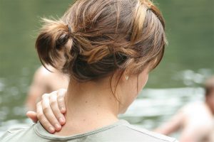
Neck Pain
Chiropractic and neck pain. Consulting a chiropractor for neck pain might seem obvious to a patient who has experienced chiropractic and who associates an adjustment with the release of tension, increased motion, and pain relief.
+ See More +
For others, chiropractic treatment of the neck brings to mind a different picture. What could be the benefit of “popping” the bones in the neck? When family doctors are asked about neck manipulation, they often visualize an abrupt twisting of the neck and are apt to sound a note of alarm when a patient seeks chiropractic care for a cervical pain and injury. However chiropractic has shown to be very effective in treating neck pain and injury. Similar to medical doctors, chiropractors use diagnostic indicators to differentiate types of neck problems. How these indicators are used and what occurs during a cervical adjustment will help to increase awareness of the indications and limitations of chiropractic care for neck conditions. Chiropractic offers a unique and valuable mechanical approach to a wide range of cervical problems. Chiropractors do not “over twist” the neck or perform painful maneuvers. Manipulation or adjusting as performed by chiropractors is a precise therapy whose aim is to return motion to restricted spinal joints and to improve the overall mechanics of the spine.
Torticollis
Torticollis, also known as cervical lock syndrome or wry neck, presents with a sudden onset of neck pain resulting with head and neck locked to one side. You may be awakened from sleep by the pain or when you wake up in the morning, you find that you have a stiff and sore neck. The cause could include a proceeding episode of intense activity, sitting in an awkward position for a period of time, sleeping with your neck exposed to an open window or even an air-conditioner. Occasionally you may have had a sore throat or an infection of the pharynx that can precipitate the cause of the stiff and sore neck. Movement of your neck is generally very difficult due to the pain and spasm.
+ See More +
Chiropractic care can be highly effective in unlocking the joints and relaxing the muscles of the neck. Modalities such as moist heat, massage, muscle stimulation, infrared light therapy and ultrasound will also speed the reduction of the spasms. With the proper treatment and within the right time frames this condition responds quickly and favorably. The longer this condition is allowed to persist, the longer the response time will be. Children and infants respond well and usually get their pain and dysfunction resolved with one to two weeks. Adults also respond well but they usually wait longer to seek care and therefore response time may be extended. Do not wait until your problem gets worse. Underlying causes need to be identified so the correct treatment can be started or to establish if the problem is more serious and needs a referral.
Whiplash
Introduction to whiplash Whiplash is a term that describes injury to the neck that occurs as a result of a motor vehicle or car accident. The most common type of car accident is the rear impact, and most typically, the occupant in the vehicle that gets “rear-ended” (hit from behind) is at the greatest risk of injury, including whiplash.
+ See More +
Until recently, the reason for the extent of whiplash injury was poorly understood. In addition, due to the legal and insurance issues, the veracity of complaints of neck pain and other symptoms by people who suffer from whiplash is commonly viewed as suspect. However, recent research has helped clarify why occupants struck from behind experience more extensive whiplash injuries than those in other types of crashes. This new information is important for the physician treating these whiplash problems, as it impacts the physician’s case management strategy. In fact, whiplash injuries can be quite complex and may include a variety of related problems, such as: Joint dysfunction Disc herniation Faulty movement patterns Chronic pain Cognitive and higher center dysfunction
How does whiplash occur?
When one motor vehicle strikes another from behind, certain forces are transmitted from the striking vehicle to the struck vehicle. These forces are then transmitted to the occupant(s) of the struck vehicle where they have the potential to cause whiplash injury. Recent research, both in the Biomechanics Laboratory at Yale University in New Haven(1), and in live crash tests using human volunteers(2), has shed new light on the contortions the cervical spine (neck) undergoes as a result of impact and culminating in whiplash. Shortly after impact (about 150 milliseconds), the cervical spine undergoes what is called an S-shaped curve. In this configuration, the cervical spine, rather than simply being curved to the front in a normal C-shape, as it would normally be at rest, takes on an altered shape: The lower part of the cervical spine moves into extension (bent backward) The upper part of the cervical spine moves into flexion (bent forward) When this whiplash occurs, the lower part of the cervical spine moves well beyond its normal range of motion, causing the potential for injury to the ligaments and discs in that area. The upper part of the cervical spine also moves beyond its normal range of motion, but to a lesser extent. There is an inherent stabilization response in the cervical spine that helps protect it from potential whiplash injury: The nervous system detects the presence of the impact; and The muscles of the cervical spine, under the direction of the nervous system, contract quickly to try to minimize the affects of the impact on the ligaments and discs. If this stabilization response is working efficiently, there is a greater likelihood of protection against whiplash with less potential for whiplash injury. But if the response is inefficient, whiplash injury is more likely. Factors affecting the whiplash injury There are several factors that affect the efficiency of the stabilization response during whiplash, some of which are within our capacity to control, others of which are not. These include: Posture at impact Overall physical condition Awareness of coming impact Gender Others
How posture at impact affects a whiplash injury
The posture in which a person is sitting at the moment of impact helps determine the efficiency of the stabilization response that will affect the severity of the whiplash injury. Sitting in a correct posture promotes an efficient stabilization response(3). Sitting in a poor posture, particularly a “slumped” type posture, promotes an inefficient stabilization response. How overall physical condition affects a whiplash injury The better conditioned the body is in general, the more efficient the stabilization response will be. This particularly relates to the condition of the nervous system, as a well-functioning nervous system is essential to a proper stabilization response. How awareness of coming impact affects a whiplash injury Perhaps the most important factor that affects the efficacy of the stabilization response in relation to whiplash is awareness of the impending impact. Scenario 1: Aware of impending impact. This person is able to automatically prepare the stabilization system to respond quickly and efficiently. Scenario 2: Unaware of the impending impact. This person cannot prepare the stabilization system, thus slowing the response and decreasing its efficiency. This person is likely to sustain greater whiplash injury than is the person who is aware. This may help explain the findings of some studies(4, 5) that have shown a passenger in a struck vehicle is likely to sustain greater whiplash injury than the driver. The driver is more likely to see the vehicle coming in the rear view mirror. How gender affects a whiplash injury Women in general are more frequently and more seriously injured by whiplash than men due to the differences in muscular bulk and the female’s smaller bony structures. These factors result in less protection of the cervical spine to the abnormal forces such as those that occur in a whiplash-type of injury. How other factors affect a whiplash injury Risk factors influencing prognosis of a whiplash injury(6): Symptoms persisting beyond 6 months. (43% failed to recover on average) Significant ligament, disc, nerve, or joint capsule injury. Delay in initiating treatment Need to resume treatment for more than one flare-up of pain. Occupant age over 65 Head restraint more than 2″ away from occupant’s head. Occupant in a small car Alcohol intoxication at time of automobile accident Pre-existing x-ray evidence of degenerative changes Prior whiplash injury Prior cervical spine fusion Patient having initial radicular (arm pain, numbness, tingling) symptoms A cervical collar used for more than 2 weeks Common misconception about whiplash injury A common misperception about whiplash injury is that if the vehicle does not sustain damage in a low speed impact, then whiplash injury to the occupant does not occur. In reality, however, low impact collisions can produce correspondingly higher dynamic loading on the occupants because the lack of crushing metal to absorb the forces results in a greater force applied to items or occupants within the vehicle(7, 8). An important point to remember is if you have been in a motor vehicle accident, you usually won’t feel the threshold of your pain or discomfort until a week or two later. Do not make the mistake of judging the severity of your condition by your pain levels. Even in a low-force crash at speeds as low as 5 mph, individuals can be subjected to whiplash. Chiropractic techniques and the chiropractor’s skill are particularly suited for correcting and relieving the otherwise debilitating effects of whiplash. At Holmes Spine & Sport Chiropractic, we are dedicated to restoring your spine to good health by restoring joint movement lost after the accident and strengthening weakened muscles through chiropractic manipulation, massage and exercise rehab. It is possible to prevent chronic or reoccurring pain from injuries suffered during whiplash. The key is not to wait, but start your chiropractic care as soon as possible.
Cervical Radiculopathy
Cervical radiculopathy is the clinical description of pain and/or neurological symptoms resulting from any type of condition that irritates a nerve in the cervical spine (neck).Cervical nerve roots, named C1 through C8, exit the cervical spine above the designated vertebral level at all levels except the last one (C8 exits below the C7 vertebra). These cervical nerves then branch out to supply muscles that enable functioning of the shoulders, arms, hands, and fingers. They also carry sensory fibers to the skin that provide sensation.When any nerve root in the cervical spine is irritated through compression or inflammation, symptoms of pain, tingling, numbness, and/or weakness can radiate anywhere along that nerve’s pathway into the shoulder, arm, and/or hand.Cervical radiculopathy symptoms most commonly appear intermittently at first—coming and going—but they could also develop suddenly or gradually.
+ See More +
There is a wide range of treatment options available for cervical radiculopathy. The treatment will depend mainly on the underlying cause of the patient’s symptoms as well as the severity of symptoms.Nonsurgical treatments will usually be tried first. If there is no improvement in symptoms after 6 to 12 weeks of treatment, then surgery might be considered. There is a wide range of treatment options available for cervical radiculopathy. The treatment will depend mainly on the underlying cause of the patient’s symptoms as well as the severity of symptoms. See Treatment for Neck Pain cage bone graft insertion.If nonsurgical treatment is unsuccessful, an anterior cervical discectomy and fusion procedure may help to relieve cervical radiculopathy symptoms. Watch: Anterior Cervical Discectomy and Fusion (ACDF) Video Nonsurgical treatments will usually be tried first. If there is no improvement in symptoms after 6 to 12 weeks of treatment, then surgery might be considered. Article continues below Nonsurgical Treatments Cervical radiculopathy nonsurgical treatments includes some combination of the following: Rest or activity modification. Oftentimes cervical radiculopathy resolves on its own, especially if the symptoms are minor. Limiting strenuous activities, like sports or lifting heavy objects, or using better posture while sitting or driving might be all that is needed. Physical therapy. An exercise and stretching routine might help relieve symptoms. A physical therapist or other certified medical professional can develop a plan that is specific for the patient.Ice and/or heat therapy. Applying an ice pack or a heated gel pack to the neck might offer pain relief for some people. For example, applying cold therapy after an activity-related flare-up of pain is often helpful in reducing inflammation and pain. Pain management with medication or injections. Various pain blockers and anti-inflammatories are available to reduce symptoms of pain. Over-the-counter (OTC) medications, such as aspirin, acetaminophen, or ibuprofen could likely be tried first. If OTC medications do not provide the patient relief, prescription-strength medications, such as muscle relaxants or opioids, could be prescribed by the doctor on a short-term basis. Another option could be an injection carefully placed with X-ray guidance to deliver medication directly into the cervical spine, such as a cervical epidural steroid injection.Manual manipulation. A chiropractor or other qualified health professional can manually adjust the cervical spine with the goal of improving mobility and providing a better healing environment. Sometimes manual manipulation is part of a physical therapy program.
Costochondritis
What is Costochondritis? Costochondritis is a condition characterized by pain and tenderness along the chest wall, normally very close to the breastbone (sternum). By definition, costochondritis is an inflammation of the juncture of the rib cartilage where the ribs attach to the sternum. While this may seem like a very simple condition, it unfortunately produces extreme pain to the point where individuals feel as if they are experiencing a heart attack or have other internal medical conditions. For this reason, it is not uncommon for individuals to go to the emergency room to rule out more serious conditions.
+ See More +
When other conditions have been ruled out, examination of the chest wall reveals isolated regions of extreme tenderness just off to the sides of the breastbone (sternum). This condition may be made worse with deep inspiration, by compression of the ribcage, or by hard coughing or sneezing. In other words, any event that either compresses or stretches the ribcage may stretch the rib cartilage, thereby producing extra pain. To make matters more complicated with this condition, costochondritis may stem simply from an inflammation of the cartilage or may be secondary to a mid-back spinal problem. For example, in a growing child who has to wear a heavy backpack, a hunching of the back or a drooping of the shoulders may be seen due to repeated postural alterations. This increased bending of the mid-back may cause compression of the ribs, causing secondary costochondritis. In this instance, chiropractic care to the thoracic spine to increase flexibility of that region, as well as mobilization techniques to the ribs, is of great benefit. At other times, simply stretching the thoracic spine to allow full movement of the rib cage brings sufficient stretch to the rib cartilage to allow the soreness to go away. Sometimes, however, localized soft tissue techniques are utilized over the rib cartilage. Transverse frictional techniques have been shown to be very effective in reducing adhesions or scar tissue that develops in cartilage. Since there is very little blood supply to the cartilage structures throughout the body, it is necessary to break up those adhesions in order for the fibers of that particular tissue to heal. Whether costochondritis comes from blunt trauma to the chest wall causing localized inflammation, a rib is injured with a twisting incident, or the thoracic spine is involved, chiropractic care normally proves very beneficial in relieving the discomfort. At other times, however, the condition may be very resistant to conservative measures and cortisone injections or anti-inflammatory medication may be needed.
Mid-Back Pain
Mid back pain is pervasive among Americans adults, but a new disturbing trend is emerging. Young children are suffering from back pain much earlier than previous generations, and the use of overweight backpacks is a contributing factor, according to the American Chiropractic Association (ACA). In fact, the U.S. Consumer Product Safety Commission reports that backpack-related injuries sent more than 7,000 people to the emergency room in 2001 alone.
+ See More +
This new back pain trend among youngsters isn’t surprising when you consider the disproportionate amounts of weight they carry in their backpacks–often slung over just one shoulder. A recent study conducted in Italy found that the average child carries a backpack that would be the equivalent of a 39-pound burden for a 176-pound man, or a 29-pound load for a 132-pound woman. Of those children carrying heavy backpacks to school, 60 percent had experienced back pain as a result. The results of these types of studies are especially important as more and more school districts–many of them in urban areas–remove lockers from the premises, forcing students to carry their books with them all day long. In fact, the problem has become so widespread that the California State Assembly recently passed legislation that would force school districts to develop ways of reducing the weight of students’ backpacks. Similar legislation is being considered in New Jersey as well. The ACA believes that limiting the backpack’s weight to no more than 10 percent of the child’s body weight and urging the use of ergonomically correct backpacks are possible solutions.
What Can You Do?
The ACA offers the following tips to help prevent the needless pain that backpack misuse could cause the students in your household. Make sure your child’s back pack weighs no more than 5 to 10 percent of his or her body weight. A heavier backpack will cause your child to bend forward in an attempt to support the weight on his or her back, rather than on the shoulders, by the straps. The backpack should never hang more than four inches below the waistline. A backpack that hangs too low increases the weight on the shoulders, causing your child to lean forward when walking. A backpack with individualized compartments helps in positioning the contents most effectively. Make sure that pointy or bulky objects are packed away from the area that will rest on your child’s back. Bigger is not necessarily better. The more room there is in a backpack, the more your child will carry-and the heavier the backpack will be. Urge your child to wear both shoulder straps. Lugging the backpack around by one strap can cause the disproportionate shift of weight to one side, leading to neck and muscle spasms, as well as low-back pain. Wide, padded straps are very important. Non-padded straps are uncomfortable, and can dig into your child’s shoulders. The shoulder straps should be adjustable so the backpack can be fitted to your child’s body. Straps that are too loose can cause the backpack to dangle uncomfortably and cause spinal misalignment and pain.
Thoracic Outlet Syndrome
The thoracic outlet of the human body is an area in which a group of nerves from the neck travel from their exit points of the neck and continues under and through the first rib and collarbone. There are also blood vessels that travel through the same pathway. The nerves, blood vessels or both can be compressed along this pathway. There are also muscles along the neck called the scalene muscles and some chest muscles, which may also cause compression of the nerves.
+ See More +
Regardless of the type of compression of the nerves and vascular tissue, you will experience symptoms into your arm. Some of the signs may be numbness and tingling as well as a diminished sensation of touch. There can also be pain and weakness. If more of the blood vessels are compressed, you may notice an increase in swelling and discoloration of the arm. This condition can mimic other conditions such as Carpal Tunnel Syndrome and/ or nerve compression at the level of the cervical spine. There are a variety of ways to diagnose and differentiate Thoracic Outlet Syndrome from other conditions. Some of them may include an electromyographic test (EMG), which can help reveal areas of nerve entrapment along the pathway of the nerve. CT scan and x-ray can also help in the diagnosis. If you are suffering from this, you may feel that your hands are going to sleep or have pins and needles in them. You also may begin to feel that your arm and hand are clumsy and weak. There may be a dull, deep ache in your arm and hand. Sometimes these symptoms will occur early in the morning and may on occasion awaken you from sleep. Prolonged sitting may also aggravate the condition, especially if these positions are accompanied by activities where you are looking down or using your hands and arms. Prolonged posture, which includes your head flexed forward, shoulders drooped or rolled forward and tightened chest muscles will also add to your symptoms
Treatment
Chiropractic manipulation . Manipulation can help reduce the joint dysfunction that may be an added component of the pain. In the initial period your chiropractor will also use to use modalities, such as heat/ice or ultrasound, to help reduce muscle spasm and lessen referred arm pain. Massage. Massage can treat active trigger points in the muscle that are producing pain and spasm that could be closing the thoracic outlet Spinal Decompression or Cervical Traction. Traction on the head can help reduce pressure from the thoracic outlet. It does not work for everyone but is easy to do, and if effective the patient can use a home traction device. Physical therapy / exercise rehab . Poor posture can decrease the thoracic out let so strengthening core muscles will improve posture opening up the thoracic outlet. Activity modification. Some types of activities may tend to aggravated thoracic outlet syndrome. Decreasing or eliminating these activities sill help reduce the pain associated with thoracic outlet syndrome. Surgical – on rare occasions surgical intervention is used to try and open up the outlet.
Coccydynia
Introduction
Coccydynia, or tailbone pain, is a fairly poorly understood condition that can cause persistent low back pain. It is felt as a localized pain at the very bottom of the spine (the coccyx) and will generally feel worse when sitting. The condition is much more common in women than men. It is usually caused by local trauma (a fall) or giving birth. On rare occasions, an infection or tumor can also cause pain in the coccyx. Local conservative treatments usually suffice to control or alleviate the pain. Rarely, surgical removal of the coccyx may be necessary if local conservative treatments are not effective in relieving the pain. Causes of coccydynia
+ See More +
The coccyx is the very bottom portion of the spine. It represents a vestigial tail (hence the common term “tailbone”) and consists of four or more very small bones fused together. The coccyx articulates with the sacrum through a vestigial disc, and is also connected to the sacrum with ligaments It is not clearly understood which portions of the anatomy can cause coccyx pain. Either the ligaments or the vestigial disc may be a cause of pain and, rarely, a primary bone tumor or soft tissue tumor can cause pain. It is thought that the condition is more common in women because: In women the coccyx is rotated and faces backward, which makes it more susceptible to trauma. Women have a broader pelvis, which means that sitting places pressure not only on their ischial tuberosities (“butt bone”) but also on the coccyx. (Men tend to sit only on their ischial tuberosities without a lot of pressure applied to the coccyx). Childbirth is a common cause of the condition. The two most common causes of coccydynia are: Local trauma. A fall on the tailbone can inflame the ligaments or injure the coccygeal attachment to the sacrum. Childbirth. During delivery, the baby’s head rides over the top of the coccyx and can injure the same structures.
Treatments
Patience is very important, since it often takes many weeks, or even months, for the pain to subside. Treatments for coccydynia: If the pain is persistent or severe, additional conservative treatments may include: Chiropracticmanipulations can provide relief. Stretching the ligaments attached to the coccyx can be helpful. Modalities with ultrasound, light/laser, electrostimulation can also be helpful. A donut-shaped pillow to help take pressure off the coccyx when sitting. Home icing. A local injection of a numbing agent (lidocaine) and steroid (to decrease inflammation in the area) can provide some relief. Anti-inflammatory medications
Facet Joint Arthritis
Osteoarthritis (degenerative arthritis) can cause breakdown of cartilage between the facet joints. When the joints move, the lack of the cartilage causes pain as well as loss of motion and stiffness. The facet joints are located in the back portion (posterior) of the spine. The joints combine with the disc space to create a three-joint complex at each vertebral level. The facet joint consists of two opposing bony surfaces with cartilage between them and a capsule around it that produces fluid. The combination of the cartilage and the fluid allows the joint to move with little friction. However, facet joint arthritis causes the cartilage to breakdown and the joint movement is associated with more friction. The patient loses motion and as they get stiffer they have more back pain.
+ See More +
Low back pain from osteoarthritis Typically, the low back pain is most pronounced first thing in the morning. Throughout the day, normal movement causes fluid to build up in the joint and it becomes better lubricated, which decreases the pain. Later in the day the pain typically becomes worse again as more stress is applied across the joint.
Treatments
Conservative treatments that concentrate on maintaining motion in the back are most effective for relieving the pain. Chiropractic manipulation : manipulations of the involved joints to increase range of motion. Exercise Rehab: Stretching exercises for the hamstring muscles, hip joints, and the back can usually serve to prevent the pain from getting worse. Aqua Aerobics – Water therapy can be also be helpful since the joints are unweighted in the water and do not generate as much pain when being moved. Medications: Acetaminophen is an effective and relatively safe non-prescription medication to help alleviate the pain, and some patients find NSAID’s (including Cox-2 inhibitors) to be helpful. Ask our medical for advice on any medications and their side effects.
Failed Back Surgery
What it is and how to avoid it
Failed back surgery introduction Failed back surgery syndrome describe the condition of patients who have not had a successful result with back surgery or spine surgery. There are many reasons that a surgery may or may not work, and even with the best surgeon and for the best indications; spine surgery is no more than 95% predictive of a successful result. Spine surgery is only basically able to accomplish two things: Decompressing a nerve root that is pinched, or Stabilizing a painful joint Unfortunately, back surgery or spine surgery cannot literally cut out a patient’s pain. It is only able to change anatomy, and an anatomical lesion (injury) that is a probable cause of back pain. By far the number one reason back surgery is not effective is because the lesion that was operated on is not in fact the cause of the patient’s pain.
+ See More +
Some types of back surgery are far more predictable in terms of alleviating a patient’s symptoms than others. For instance, A discectomy (or microdiscectomy) for a lumbar disc herniation that is causing leg pain is a very predictable operation. However, a discectomy for a lumbar disc herniation that is causing lower back pain is far less likely to be successful. A spine fusion for spinal instability (e.g. spondylolisthesis) is a relatively predictable operation. However, a spine fusion for multi-level lumbar degenerative disc disease is far less likely to be successful in reducing a patient’s pain. In addition to the above-mentioned cause of failed back surgery syndrome, there are several other potential causes of a failed surgery, or continued pain after surgery: Fusion surgery considerations (such as failure to fuse and/or implant failure, or a transfer lesion to another level after a spine fusion, when the next level degenerates and becomes a pain generator) Lumbar decompression back surgery considerations (such as recurrent stenosis or disc herniation, inadequate decompression of a nerve root, preoperative nerve damage that does not heal after a decompressive surgery, or nerve damage that occurs during the surgery) Scar tissue considerations (scarring tissue can wrap itself around the nerve becoming a constant source of pain) Therefore, the best way to avoid a spine surgery that leads to an unsuccessful result is to stick to operations that have a high degree of success and to make sure that an anatomic lesion that is amenable to surgical correction is identified preoperatively. Research indicates that chiropractic manipulation combined with specific strength and stabilizing rehabilitation exercises have a higher chance of easing and correcting your cause of pain than medication or physical therapy alone. Holmes Spine & Sport also utilizes an active spinal decompression table to release nerve pressure and help heal herniated disc and sciatica pain. Chiropractic and other conservative care treatments can still be effective even when surgery has failed to relieve the pain . Holmes Spine & Sport Chiropractic provides a number of conservative treatments which makes it easier to get the care you need. We also work with your family physician and orthopedic surgeon.
Low-Back Pain
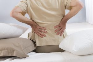
The lumbar spine, or low back, is a remarkably well-engineered structure of interconnecting bones, joints, nerves, ligaments, and muscles all working together to provide support, strength, and flexibility. However, this complex structure also leaves the low back susceptible to injury and pain.To help understand this complicated topic, this article presents a model for understanding symptoms, physical findings, imaging studies and injection techniques to come to a precise diagnosis. Once an accurate diagnosis of the cause of the lower back pain is attained, treatment options can be selected based on today’s best medical practices.
+ See More +
The Lumbar Spine, What Can Go Wrong
The low back supports the weight of the upper body and provides mobility for everyday motions such as bending and twisting. Muscles in the low back are responsible for flexing and rotating the hips while walking, as well as supporting the spinal column. Nerves in the low back supply sensation and power the muscles in the pelvis, legs, and feet. Most acute low back pain results from injury to the muscles, ligaments, joints, or discs. The body also reacts to injury by mobilizing an inflammatory healing response. While inflammation sounds minor, it can cause severe pain. There is a significant overlap of nerve supply to many of the discs, muscles, ligaments, and other spinal structures, and it can be difficult for the brain to accurately sense which is the cause of the pain. For example, a degenerated or torn lumbar disc can feel the same as a pulled muscle – both creating inflammation and painful muscle spasm in the same area. Muscles and ligaments heal rapidly, while a torn disc may or may not. The time course of pain helps determine the cause.
Lower Back Pain Symptoms
Low back pain might begin as acute due to an injury, but can become chronic. Managing pain appropriately at an early stage can help limit symptoms in both time and severity. Identifying the symptoms and getting a diagnosis that pinpoints the underlying cause of the pain is the first step in obtaining effective pain relief.
Early Treatments for Lower Back Pain
Many treatment options for low back pain can be tailored to an individual patient’s needs. Treatments include care administered at home, medicinal remedies, alternative care, or even surgery.Depending on the patient’s diagnosis, some treatments may be more effective than others. Many people find that a combination of treatments is best. Self-Care for Low Back Pain Basic remedies applied at home can be effective for treating mild or acute pain from muscle strain, as well as reducing the effects of chronic, severe pain. Self-care is administered by the individual and can easily be adjusted. These methods include: Short rest period. Many episodes of lower back pain can be improved by briefly avoiding strenuous activity. It is not advised to rest for more than a few days, as too much inactivity can make healing more difficult. Activity modification. One variant of resting is to stay active but avoid activities and positions that aggravate the pain. For example, if long periods of sitting in a car or at a desk make the pain worse, then set a timer to get up every 20 minutes and walk around or gently stretch. If standing makes the pain worse, avoid chores that require standing such as washing dishes at the sink. Avoiding, or minimizing, activities and positions that worsen the pain will help prevent or reduce painful back spasms and allow for a better healing environment.Heat/ice therapy. Heat from a warm bath, hot water bottle, electric heating pad, or chemical or adhesive heat wraps can relax tense muscles and improve blood flow. Increased blood flow brings nutrients and oxygen that muscles need to heal and stay healthy. If the low back is painful due to inflammation, ice or cold packs can be used to reduce swelling. It’s important to protect the skin while applying heat and ice to prevent tissue damage. Alternating heat and ice can be especially helpful when returning to activity: applying heat before activities helps relax muscles, allowing for better flexibility and mobility; applying ice after activity reduces the chances of an area becoming irritated and swollen from exercise. Over-the-counter pain medications. The most common over-the-counter (OTC) medications are aspirin (e.g. Bayer), ibuprofen (e.g. Advil), naproxen (e.g. Aleve), and acetaminophen (e.g. Tylenol). Aspirin, ibuprofen, and naproxen are anti-inflammatory medicines, which alleviate low back pain caused by a swollen nerves or muscles. Acetaminophen works by interfering with pain signals sent to the brain. Self-care treatments generally do not need guidance from a doctor, but should be used carefully and attentively. Any type of medication carries possible risks and side effects. If a patient is unsure which kinds of self-care would work best, talking to a doctor is advised. Low back pain might begin as acute due to an injury, but can become chronic. Managing pain appropriately at an early stage can help limit symptoms in both time and severity. Lumbar Degenerative Disc Disease Pain is typically divided into three categories: acute, chronic, and neuropathic. Read: Types of Back Pain: Acute Pain, Chronic Pain, and Neuropathic Pain Identifying the symptoms and getting a diagnosis that pinpoints the underlying cause of the pain is the first step in obtaining effective pain relief. See Diagnosing the Cause of Lower Back Pain Article continues below Common Symptoms of Lower Back Problems Specifically identifying and describing symptoms can help lead to a more accurate diagnosis and effective treatment plan. Low back pain is typically characterized by a combination of the following symptoms: Dull, aching pain. Pain that remains within the low back (axial pain) is usually described as dull and aching rather than burning, stinging, or sharp. This kind of pain can be accompanied by mild or severe muscle spasms, limited mobility, and aches in the hips and pelvis. Pain that travels to the buttocks, legs, and feet. Sometimes low back pain includes a sharp, stinging, tingling or numb sensation that moves down the thighs and into the low legs and feet, also called sciatica. Sciatica is caused by irritation of the sciatic nerve, and is usually only felt on one side of the body. Pain that is worse after prolonged sitting. Sitting puts pressure on the discs, causing low back pain to worsen after sitting for long periods of time. Walking and stretching can alleviate low back pain quickly, but returning to a sitting position may cause symptoms to return. Pain that feels better when changing positions. Depending on the underlying cause of pain, some positions will be more comfortable than others. For example, with spinal stenosis walking normally may be difficult and painful, but leaning forward onto something, such as a shopping cart, may reduce pain. How symptoms change with shifting positions can help identify the source of pain. Pain that is worse after waking up and better after moving around. Many who experience low back pain report symptoms that are worse first thing in the morning. After getting up and moving around, however, symptoms are relieved. Pain in the morning is due to stiffness caused by long periods of rest, decreased blood flow with sleep, and possibly the quality of mattress and pillows used
article by Spine-Health
Lumbar Degenerative Disk
Degenerative disc disease refers to a syndrome in which a compromised disc causes low back pain. Lumbar degenerative disc disease usually starts with a torsional (twisting) injury to the lower back, such as when a person rotates to put something on a shelf or swing a golf club. However, the pain is also frequently caused by simple wear and tear on the spine. Despite its rather dramatic label, degenerative disc disease is fairly common, and it is estimated that at least 30% of people aged 30-50 years old will have some degree of disc space degeneration, although not all will have pain or ever receive a formal diagnosis. In fact, after a patient reaches 60, some level of disc degeneration is deemed to be a normal finding, not the exception.
+ See More +
Lumbar degenerative disc disease pain and symptoms Most patients with lumbar degenerative disc disease will experience low-grade continuous but tolerable pain that will occasionally flare (intensify) for a few days or more. Pain symptoms can vary, but generally are: Centered on the lower back, although it can radiate to the hips and legs Frequently worse when sitting, when the discs experience a heavier load than when patients are standing, walking or even laying down Exacerbated by certain movements, particularly bending, twisting or lifting The low back pain associated with lumbar degenerative disc disease is usually generated from one or both of two sources: Inflammation, as the proteins in the disc space irritate the surrounding nerves, and/or Abnormal micro-motion instability, when the outer rings of the disc – the annulus fibrous – are worn down and cannot absorb stress on the spine effectively, resulting in movement along the vertebral segment Excessive micro-motion, combined with the inflammatory proteins, can produce ongoing low back pain Fortunately, over time the pain from lumbar degenerative disc disease usually decreases, rather than becoming progressively worse. This is because a fully degenerated disc no longer has any inflammatory proteins (that can cause pain) and usually collapses into a stable position, eliminating the micro-motion that generates the pain.
Diagnosis
Following a review of the patient’s history and a physical examination, a formal diagnosis of lumbar degenerative disc disease can be confirmed with x-rays, ct scan, or magnetic resonance imaging (MRI). Findings that are closely linked to a painful disc include disc space collapse of greater than 50% and cartilaginous endplate erosion.
Treatment
For most people, degenerative disc disease can be successfully treated with conservative (meaning non-surgical) care consisting of exercise, chiropractic, massage and ice and heat applications. Medication can also control inflammation and pain, however the effects that NSAIDS have on stomach linings and kidneys should make a person think twice for relying on medication as a long term treatment of degenerative disc disease. Surgery is only considered when patients have not achieved relief from conservative care and/or are significantly constrained in performing everyday activities.
Non-surgical treatment for degenerative disc disease
Chiropractic manipulation can relieve low back pain by taking pressure off sensitive nerves or tissue, increasing range of motion, restoring blood flow, reducing muscle tension, and, like more active exercise, promoting the release of endorphins within the body to act as natural painkillers Applying heat to stiff muscles or joints to increase flexibility and range of motion, or using ice packs to cool down sore muscles or numb the area where painful flares are concentrated. Medications such as non-steroidal anti-inflammatories may be used to manage intense pain episodes on a short-term basis, and some patients may benefit from an epidural steroid injection. Not all medications are right for all patients, and patients will need to discuss side effects and possible factors that would preclude taking them with their physician. An exercise program is essential to relieving the pain of lumbar degenerative disc disease and should have several components, including: Hamstring stretching, since tightness in these muscles can increase the stress on the back and the pain caused by a degenerative disc A strengthening exercise program, such as Dynamic Lumbar Stabilization exercises, where patients are taught to find their ‘natural spine’, the position in which they feel most comfortable, and to maintain that position Low-impact aerobic conditioning (such as walking, swimming, biking) to ensure adequate flow of nutrients and blood to spine structures, and relieve pressure on the discs Traction Decompression Therapy and relieve pressure on sensitive nerve structures At Holmes Spine & Sport Chiropractic we provide a number of conservative treatments under one roof to treat your lumbar disc degeneration. We also work with your family physician or orthopedic surgeon to assist in getting you any medications or consultations you may need.
Lumbar Disc Herniation
Introduction
A common cause of low back and leg pain is a ruptured or herniated disc. Symptoms may include dull or sharp pain, muscle spasm or cramping, sciatica, and leg weakness or loss of leg function. Sneezing, coughing, or bending usually intensifies the pain. Rarely bowel or bladder control is lost, and if this occurs, seek medical attention at once.
+ See More +
Sciatica is a symptom frequently associated with a lumbar herniated disc. Pressure on one or several nerves that contribute to the sciatic nerve can cause pain, burning, tingling, and numbness that extends from the buttock into the leg and sometimes into the foot. Usually one side (left or right) is affected. Anatomy – Normal Lumbar Disc In between each of the five lumbar vertebrae (bones) is a disc, a tough fibrous shock-absorbing pad. Endplates line the ends of each vertebra and help hold individual discs in place. Each disc contains a tire-like outer band (called the annulus fibrosus) that encases a gel-like substance (called the nucleus pulposus). Nerve roots exit the spinal canal through small passageways between the vertebrae and discs. Pain and other symptoms can develop when the damaged disc pushes into the spinal canal or nerve roots.
How a herniated disc causes pain
As a disc degenerates, it can herniate (the inner core extrudes) back into the spinal canal, which is known as a disc herniation (or a herniated disc). The weak spot in a disc is directly under the nerve root, and a herniated disc in this area puts direct pressure on the nerve, which in turn can cause pain to radiate all the way down the patient’s leg to the foot Approximately 90% of disc herniations will occur at L4- L5 (lumbar segments 4 and 5) or L5- S1 (lumbar segment 5 and sacral segment1), which causes pain in the L5 nerve or S1 nerve, respectively. L5 nerve impingement from a herniated disc can cause weakness in extension of the big toe and potentially in the ankle (foot drop). Numbness and pain can be felt on top of the foot, and the pain may also radiate into the rear. S1 nerve impingement from a herniated disc may cause loss of the ankle reflex and/or weakness in ankle push off (e.g. patients cannot do toe rises). Numbness and pain can radiate down to the sole or outside of the foot
Progression of disc herniation
Disc Degeneration: chemical changes associated with aging causes discs to weaken, but without a herniation. Prolapse: the form or position of the disc changes with some slight impingement into the spinal canal. Also called a bulge or protrusion. Extrusion: the gel-like nucleus pulposus breaks through the tire-like wall (annulus fibrosus) but remains within the disc. Sequestration or Sequestered Disc: the nucleus pulposus breaks through the annulus fibrosus and lies outside the disc in the spinal canal (HNP).
Treatment
Conservative treatment options for a lumbar herniated disc There are a number of non-surgical treatment options that can help alleviate the pain and hel heal a lumbar herniated disk: Chiropractic manipulation. Gentle manipulation can help reduce the joint dysfunction and disc pressure. In the initial treatment period your chiropractor may also use modalities, such as heat/ice or ultrasound, to help reduce muscle spasm and lessen referred leg pain. Massage. Massage can treat active trigger points and associated muscle tightness and spasm related to the nerve compression. Spinal Decompression or Lumbar Traction. Traction/ decompression of the lumbar spine can help reduce pressure over the nerve root. It does not work for everyone but for those that get relief it can be an effective way to treat lumbar disc herniations. Physical therapy / exercise rehab. Mckenzie exercises can be used to help reduce the pain in the leg. As the disc and nerve heal, more intense exercises can be added to stabilize the disc and joint complex. Activity modification. Some types of activities may tend to exacerbate the herniated disc pain and it is reasonable to avoid these activities to keep from irritating the nerve root. Such activities may include heavy lifting, activities that can cause increased vibration and compression to the lumbar spine (boating, snowmobile riding, running, etc.). Bracing. In some instances a lumbar brace/support belt may be recommended to help provide some rest for the lumbar spine. Medications. In addition to the anti-inflammatory medication, narcotic agents (pain killers) might be used on a temporary basis to help reduce the pain and discomfort from a lumbar herniated disc. Also, muscle relaxants or certain anti-depressants may help reduce the nerve-type pain (neuropathic pain) and help restore normal sleep patterns. For patients with severe pain from a herniated disc, oral steroids (such as Predisone or a Medrol Dose Pak) may give even better pain relief. However, these medications can only be used for a short period of time (one week). Injections. Epidural steroid injections or selective nerve root blocks can be helpful to reduce inflammation in cases of severe pain from a lumbar herniated disc, and can be very effective if accompanied by a comprehensive rehabilitation program that may involve a number of the above conservative treatments. Spine surgery for a lumbar herniated disc Most episodes of leg pain due to a lumbar herniated disc will resolve over a period of weeks to months. However, if the pain and disability is not improving or is worsening spine surgery may be a reasonable option.
Summary of lumbar herniated disc treatment options
Lumbar herniated disc typically respond well to conservative treatments. For the few cases that don’t respond well surgery may be your only option At Holmes Spine & Sport Chiropractic we provide a number of conservative treatments under one roof to treat your lumbar disc herniation. We also work with your family physician or orthopedic surgeon to assist in getting you any medications or consultations you may need.
Lumbar Strain
Lumbar strain: A stretching injury to the ligaments, tendons, and/or muscles of the low back. The stretching incident results in microscopic tears of varying degrees in these tissues. Lumbar strain is one of the most common causes of low back pain. The injury can occur because of overuse, improper use, or trauma. It is classified as “acute” if it has been present for days to weeks. If the strain lasts longer than 3 months, it is referred to as “chronic.”
+ See More +
Symptoms
Lumbar strain most often occurs in persons in their forties, but can happen at any age. The condition is characterized by localized discomfort in the low back area with onset after an event that mechanically stressed the lumbar tissues. The diagnosis of lumbar strain is based on the history of injury, the location of the pain, and exclusion of nervous system injury. Usually, x-ray testing is only helpful to exclude bone abnormalities.
Treatment
The treatment of lumbar strain consists of resting the back (to avoid re-injury), medications to relieve pain and muscle spasm, local heat applications, massage, and eventual (after the acute episode resolves) reconditioning exercises to strengthen the low back and abdominal muscles. Long periods of inactivity in bed are no longer promoted as this treatment may actually slow recovery. Spinal manipulation for periods of up to 1 month has been found helpful in some patients that do not have signs of nerve irritation. Future injury is avoided by using back protection techniques during activities and support devices as needed at home or work.
article by MedicineNet.com
Piriformis Syndrome
Piriformis syndrome and sciatica pain Piriformis syndrome is a condition in which the piriformis muscle irritates the sciatic nerve and causes pain in the rear and may cause pain along the back of the leg and into the foot (similar to sciatica pain). Piriformis syndrome is most common among women, and is thought to be common among active individuals (such as runners and walkers).
+ See More +
While there is some controversy in the medical community, many health professionals believe that an accurate diagnosis and comprehensive management approach are critical to alleviate the sciatica type of pain caused by piriformis syndrome.
What is the piriformis muscle?
The piriformis muscle is a small muscle located deep in the rear (deep to the gluteus maximus). The piriformis muscle: Starts at the lower spine and connects to the upper surface of each femur (thighbone). Functions to assist in rotating the hip. Runs horizontally, with the sciatic nerve running vertically directly beneath it. Piriformis syndrome can develop when the piriformis muscle becomes tight or spasms and places pressure on the sciatic nerve that runs beneath it. The pressure on the sciatic nerve can cause low back pain and/or pain that radiates to the rear and down the leg (similar to sciatica pain). From a technical standpoint, piriformis syndrome does not cause true sciatica (as sciatica is usually defined as a radiculopathy, or compression of a nerve root as it exits the spine). However, just like sciatica, piriformis syndrome can cause pain, numbness and tingling along the sciatic nerve, which runs down the back of the leg and into the foot.
Diagnosis of piriformis syndrome
There is no simple diagnostic test for piriformis syndrome causing irritation of the sciatic nerve. The condition is primarily diagnosed on the basis of the patient’s symptoms and on a physical exam.
Symptoms of piriformis syndrome
Most commonly, patients describe acute tenderness in the rear and sciatica-like pain down the back of the leg. Typical symptoms of piriformis syndrome may include: A dull ache in the mid-rear Pain down the back of the leg (a radiculopathy or sciatica) Pain when walking up stairs or inclines Increased pain after prolonged sitting Symptoms of piriformis syndrome often become worse after prolonged sitting, walking or running, and may feel better after lying down on the back. Physical exam to diagnose piriformis syndrome The physical exam will include examination of the hip and legs to see if movement causes increased low back pain or leg pain (sciatica pain). Typically, motion of the hip will recreate the pain. X-rays and other spinal imaging studies cannot detect if the sciatic nerve is being irritated at the piriformis muscle. However, diagnostic tests (such as X-rays, MRI and nerve conduction tests) may be conducted to exclude other conditions that can cause similar symptoms to piriformis syndrome (such as a disc herniation). Comprehensive management of piriformis syndrome and sciatica pain Depending on the severity of the patient’s sciatica-type pain and other symptoms, a number of treatment options may be recommended by a health care professional. A comprehensive approach to managing pain along the sciatic nerve from piriformis syndrome may include a combination of: Chiropractic manipulation of the lumbar and sacral joints can restore proper motion to the spine and create an inhibitory reflex to shut down a spasm of the piriformis. Stretching exercise for piriformis syndrome A number of stretching exercises for the piriformis, hamstrings and hip extensors may help decrease the painful symptoms along the sciatic nerve and return the patient’s range of motion. Ice for piriformis syndrome At the onset of pain, lie in a comfortable position on your stomach and place an ice pack on the painful area for approximately 15 minutes. Repeat as needed every 2 to 4 hours. If specific activities are usually followed by increased pain, it may be a good idea to apply ice immediately following the activity. Range of motion exercises A physical therapist can develop a customized program of stretching and range of motion exercises to help stretch the muscle and decrease spasm. Deep Massage Deep massage (manual release) by a physical therapist is thought to enhance healing by increasing blood flow to the area and decreasing muscle spasm. Heat /Ice Some people find it helpful to use either ice or heat or to alternate cold with heat. Medications for sciatica pain Since most episodes of pain include some type of inflammation, non-steroidal anti-inflammatory medications (NSAID’s) (such as ibuprofen or naproxen) may help decrease inflammation in the affected area. Injections for sciatica pain and piriformis syndrome For severe sciatica pain, a local anesthetic and corticosteroid may be injected in directly into the piriformis muscle to help decrease the spasm and help alleviate the sciatica pain. The purpose of an injection is usually to decrease acute pain to enable progress in physical therapy. For persistent piriformis spasm that is resistant to anesthetic/corticosteroid injections, an injection of botulinum toxin (a muscle weakening agent) may be useful. Electrotherapy and ultrasound for piriformis syndrome The application of electrical stimulation and ultrasound to the rear with can help to block pain and reduce muscle spasm related to piriformis syndrome.
Sacroiliac Dysfunction
What is sacroiliac joint dysfunction?
Dysfunction in the sacroiliac joint is thought to cause low back and/ or leg pain. The pain can be similar to pain caused by a lumbar disc herniation. This condition is generally more common in young and middle age women. The anatomical source of sacroiliac joint pain The sacroiliac joint lies next to the spine and connects the sacrum (the triangular bone at the bottom of the spine) with the pelvis (iliac crest). The joint:
+ See More +
Is small and very strong Transmits all the forces of the upper body to the pelvis (hips) and legs Acts as a shock-absorbing structure Has limited motion While it is not clear how the pain is caused, it is thought that an alteration in the normal joint motion may be the culprit that causes sacroiliac joint pain. This source of pain can be caused by either: Too much movement — hypermobility or instability, or Too little movement — hypomobility or fixation. The pain is typically felt on one side of the low back or buttocks, and can radiate down the leg. The pain usually remains above the knee, but at times pain can extend to the ankle or foot.
Diagnosis
Accurately diagnosing sacroiliac joint dysfunction can be difficult. The symptoms mimic other common conditions, such as disc herniation and radiculopathy (pain along the sciatic nerve that radiates down the leg). A diagnosis is usually arrived at through physical examination: Physical examination to determine the source of pain In physical examination, the doctor may try to determine if the sacroiliac joint is the cause of pain through movement of the joint. If the movement recreates the patient’s pain, and no other cause of pain has been found (such as a disc herniation on an MRI scan), the sacroiliac joint may be the cause of the pain. There are several orthopedic provocative tests that can be used in attempt to reproduce the symptoms associated with sacroiliac joint dysfunction. As a rule, several positive tests that reproduce pain specifically located at the sacroiliac joint improves the probability of the diagnosis of sacroiliac joint dysfunction.
Treatment
Treatment for sacroiliac joint dysfunction is usually conservative (non-surgical) and focuses on restoring normal motion in the joint. One of the most effective ways to do this is through the chiropractic manipulation of the sacroiliac joint. Injections, exercise and NSAIDS may also may be used to treat sacroiliac pain.
Sciatica
What you need to know about sciatica
Understanding sciatica Low back pain and/or leg pain that usually travels down the large sciatic nerve, from the lower back down the back of each leg, is generally referred to as sciatica and is fairly common. This pain can be caused when a nerve root in the lower spine that helps form the sciatic nerve is pinched or irritated.
+ See More +
Sciatica is usually caused by pressure on the sciatic nerve from a herniated disc (also referred to as a ruptured disc, pinched nerve, slipped disk, etc.) in the lumbar spine. Sciatica occurs most frequently in people between 30 and 50 years of age. Often a particular event or injury does not cause sciatica, but rather it may develop as a result of general wear and tear on the structures of the lower spine. The vast majority of people who experience sciatica get better with time (usually a few weeks or months) and find pain relief and correction with non-surgical treatments
Understanding sciatica pain
For some people, the pain from sciatica can be severe and debilitating. For others, the pain might be infrequent and irritating, but has the potential to get worse. Usually, sciatica only affects one side of the lower body, and the pain often radiates from the lower back all the way through the back of the thigh and down through the leg. Depending on where the sciatic nerve is affected, the pain may also radiate to the foot or toes. One or more of the following sensations may occur as a result of sciatica: Pain in the rear or leg that is worse when sitting Burning or tingling down the leg Weakness, numbness or difficulty moving the leg or foot A constant pain on one side of the rear A shooting pain that makes it difficult to stand up Low back pain may be present along with the leg pain, but usually the low back pain is less severe than the leg pain While sciatica can be very painful, it is rare that permanent nerve damage (tissue damage) will result. Most sciatica pain syndromes result from inflammation and will get better within two weeks to a few months. Also, because the spinal cord is not present in the lower (lumbar) spine, a herniated disc in this area of the anatomy does not present a danger of paralysis. Symptoms that may constitute a medical emergency include progressive weakness in the leg or bladder/bowel incontinence. Patients with these symptoms may have cauda equina syndrome and should seek immediate medical attention. Any condition that causes irritation or impingement on the sciatic nerve can cause the pain associated with sciatica. The most common cause is a lumbar herniated disc. Other common causes of sciatica include lumbar spinal stenosis, degenerative disc disease, or spondylolisthesis, sacro-iliac joint dsfunction and piriformis syndrome. Sciatica symptoms and causes Back problems and the sciatic nerve The sciatic nerve is the largest nerve in the body and is composed of individual nerve roots that combine to form the “sciatic nerve”. It starts in the low back at lumbar segment 3 (L3). The sciatic nerve roots run through the bony canal in the spine, and at each level in the lower back a pair of nerve roots exits from the spine and then comes together to form the large sciatic nerve that runs all the way down the back of each leg. Portions of the sciatic nerve then branch out in each leg to innervate certain parts of the leg (e.g. the calf, the foot, the toes). The nerve roots that originate in the lower back are named for the upper vertebral body that they run between (for example, the nerve that exits at L4-L5 in the spine is named L4). The nerve passing to the next level runs over a weak spot in the disc space, which is the reason discs tend to herniate (extrude) right under the sciatic nerve root and can cause sciatica. The sciatica symptoms (e.g., low back pain, leg pain, numbness, tingling, weakness) are different depending on where the pressure on the nerve occurs. For example, a lumbar segment 5 (L5) nerve impingement can cause weakness in extension of the big toe and potentially in the ankle (foot drop) . Treatment of sciatica requires a diagnosis of the underlying cause of the sciatica pain.
Symptoms
Common low back problems and other spinal conditions that can cause sciatica – pain along the sciatic nerve – include: Lumbar herniated disc. A herniated disc occurs when the soft inner core of the disc (nucleus pulposus) extrudes through the fibrous outer core (annulus) of the disc and the bulge places pressure on the contiguous nerve root as it exits the spine. In general, it is thought that a sudden twisting motion or injury can lead to an eventual disc herniation and sciatica. However, most discs weaken due to repetitive stress and the final result is a herniation. A herniated disc is sometimes referred to as a slipped disk, ruptured disk, bulging disc, protruding disc, or a pinched nerve. * See also Lumbar disc herniation Lumbar spinal stenosis. This condition commonly causes sciatica due to a narrowing of the spinal canal. It is more common in adults over age 60, and typically results from a combination of one or more of the following: enlarged facet joints, overgrowth of soft tissue and a bulging disc placing pressure on the nerve roots as they exit the spine. * See also Lumbar spinal stenosis Degenerative disc disease. While disc degeneration is a natural process that occurs with aging, in some cases one or more degenerated discs can also irritate a nerve root and cause sciatica. Degenerative disc disease is diagnosed when a weakened disc results in excessive micro-motion at the corresponding vertebral level and inflammatory proteins from inside the disc become exposed and irritate the area (including the nerve roots). The term “degenerative disc disease” is an unfortunate one as it is a process, not a disease. * See also Degenerative disc disease Spondylolisthesis. This condition is not that uncommon in adults (approximately 5% to 7% of adults are thought to have it), but it only rarely causes back pain or sciatica pain. Spondylolisthesis occurs when a small stress fracture (most often at the fifth segment) allows the L5 vertebral body to slip forward on the S1 vertebral body. Caused by a combination of disc space collapse, the fracture, and the vertebral body slipping forward, the L5 nerve can get pinched as it exits the spine. * See also spondylolisthesis Piriformis syndrome. The sciatic nerve can also get irritated as it runs under the piriformis muscle in the rear. If the piriformis muscle irritates or pinches a root that comprises the sciatic nerve, it can cause sciatica-type pain. This is not a true radiculopathy, but the pain can feel the same as sciatica caused by a nerve irritation. * See also Piriformis syndrome—another irritation to the sciatic nerve Sacroiliac joint dysfunction. Irritation of the sacroiliac joint at the bottom of the spine can also irritate the L5 nerve, which lies on top of it, and cause sciatica-type pain. This is not a true radiculopathy, but the pain can feel the same as sciatica caused by a nerve irritation. * See also What is sacroiliac joint dysfunction?
Sciatica treatments
The good news for patients is that sciatica usually will get better over time, and the healing process usually only takes a few days or weeks. Overall, the vast majority of episodes of sciatica pain heal within a six to twelve week time span. However, occasional flare-ups of sciatic nerve pain may be an indication of a condition that should be managed so that it does not get worse over time. For most, chiropractic and regular exercise will go a long way to remedying the situation. Sciatica medical treatments During an episode of sciatica, there are a number of conservative care (meaning non-surgical) options available to help alleviate the sciatic pain and discomfort. Manual manipulation Chiropractic spinal adjustments and manual manipulation are focused on providing better spinal column alignment, which in turn should help to address a number of underlying conditions that can cause sciatic nerve pain. Chiropractic manipulation addresses the cause of the sciatic pain and can provide lasting relief. Heat/ice for sciatica For acute sciatica pain, heat and/or ice packs are most readily available and can help alleviate the pain, especially in the acute phase. Usually ice or heat is applied for approximately 20 minutes, and repeated every two hours. Most people use ice first, but some people find more relief with heat. The two may be alternated to help with sciatica pain relief. Massage therapy Certain forms of massage therapy have been shown to have a number of benefits for back pain, including increased blood circulation, muscle relaxation, and release of endorphins (the body’s natural pain relievers). Spinal Decompression or Lumbar Traction Traction on the lumbar spine can help reduce pressure over the nerve root. It does not work for everyone but for those that get relief it can be an effective way to treat sciatica. Medications for sciatica Over-the-counter or prescription medications may also be helpful in relieving sciatica. Non-steroidal anti-inflammatory drugs (such as ibuprofen, naproxen, or COX-2 inhibitors), or oral steroids can be helpful in reducing the inflammation and pain associated with sciatica. Please talk to your medical doctor regarding treatment with drugs and their associated side effects Epidural steroid injections for sciatica If the sciatica pain is severe, an epidural steroid injection can be performed to reduce the inflammation. An epidural injection is different from oral medications because it injects steroids directly to the painful area around the sciatic nerve to help decrease the inflammation that may be causing the pain. While the effects tend to be temporary (providing pain relief for as little as one week up to a year), and it does not work for everyone, an epidural steroid injection can be effective in providing relief from an acute episode of sciatic pain. Importantly, it can provide sufficient relief to allow a patient to progress with a conditioning and exercise program. Physical therapy and exercise When the sciatica pain is at its worst, patients may need to rest for a day or two, but resting for longer periods of time is usually not advisable. In fact, inactivity will usually make the sciatic pain worse. This is because regular movement and exercise is necessary to nourish the various structures in the low back and encourage the strength needed to support the low back. Many sciatica exercises focus on strengthening the abdominal and back muscles in order to give more support for the back. Stretching exercises for sciatica target muscles that cause pain when they are tight and inflexible. When patients engage in a regular program of gentle strengthening and stretching exercises, they can recover more quickly from a flare up of sciatica and can help to prevent future episodes of pain. Low impact aerobic exercise, such as walking or swimming (or pool therapy) is also usually a component of recovery, as aerobic activity encourages the exchange of fluids and nutrients to help create a better healing environment. Aerobic conditioning also has the added benefit of releasing endorphins, the body’s natural pain killers, which is a natural way to alleviate sciatic pain. Lumbar laminectomy (open decompression) for sciatica Lumbar spinal stenosis often causes sciatica pain that waxes and wanes over many years. Surgery may be offered as an option if the patient’s activity tolerance falls to an unacceptable level. Again, surgery is elective and need only be considered for those patients who have not gotten relief from the sciatica after pursuing non-surgical treatments. The patient’s general health may also be a consideration. After a lumbar laminectomy (open decompression), approximately 70% to 80% of patients typically experience relief from their sciatic nerve pain. It is important to note that the decision whether or not to pursue surgical or non-surgical remedies for sciatica is almost always the patient’s decision. There are many considerations for the patient to take into account when deciding whether or not to have surgery. At Holmes Spine & Sport Chiropractic we provide a number of conservative treatments to treat your sciatic pain. We also work with your family physician or orthopedic surgeon to assist in getting you any medications you may need.
Spondylolisthesis
What is spondylolisthesis?
Spondylolisthesis is a condition in which one bone in your back (vertebra) slides forward over the bone below it. It most often occurs in the lower spine (lumbosacral camera.gif area). In some cases, this may lead to your spinal cord or nerve roots being squeezed. This can cause back pain and numbness or weakness in one or both legs. In rare cases, it can also lead to losing control over your bladder or bowels. See a doctor right away if you begin losing bladder or bowel control.
Sometimes when a vertebra slips out of place, you may have no symptoms at all or no symptoms until years later. Then, you may have pain in your low back or buttock. Muscles in your leg may feel tight or weak. You may even limp.
+ See More +
What causes spondylolisthesis?
The bones in your spine come together at several small joints that keep the bones lined up while still allowing them to move. Spondylolisthesis is caused by a problem with one or more of these small joints that allows one bone to move out of line.
Spondylolisthesis may be caused by any of a number of problems with the small joints in your back. You could have:
- A defective joint that you’ve had since birth (congenital).
- A joint damaged by an accident or other trauma.
- A vertebra with a stress fracture caused from overuse of the joint.
- A joint damaged by an infection or arthritis.
- Spondylolisthesis affects children and teens involved in sports. Some sports, such as gymnastics or weight lifting, can overuse back bones to the point of causing stress fractures in vertebrae, which can result in spondylolisthesis.
Older adults can develop spondylolisthesis, because wear and tear on the back leads to stress fractures. It can also occur without stress fractures when the disc and joints are worn down and slip out of place.
What are the symptoms?
Symptoms of spondylolisthesis may include:
- Back or buttock pain.
- Pain that runs from the lower back down one or both legs.
- Numbness or weakness in one or both legs.
- Difficulty walking.
- Leg, back, or buttock pain that gets worse when you bend over or twist.
- Loss of bladder or bowel control, in rare cases.
- Sometimes spondylolisthesis causes no symptoms at all.
How is spondylolisthesis diagnosed?
Your doctor will look at X-rays of your back if he or she suspects you have spondylolisthesis. X-rays will show if any of the vertebrae in your back have fractures or cracks and have slipped out of place. You could also have a CT scan or an MRI to pinpoint the damage and help guide treatment.
How is it treated?
Treatment for spondylolisthesis begins with stopping any physical activity that may have led to vertebrae damage. To help relieve pain, take nonsteroidal anti-inflammatory drugs, including ibuprofen (such as Advil) or naproxen (such as Aleve). Do not give aspirin to anyone younger than 20 because of the risk of Reye syndrome, a serious illness. Acetaminophen (such as Tylenol) can also help with pain. Be safe with medicines. Read and follow all instructions on the label.
Doctors often suggest physical therapy to build up stomach and back muscles (core strengthening). In overweight people, weight loss may also help.
When pain is extreme or bones continue to move, or if there is nerve root or spinal cord damage related to the spondylolisthesis, surgery can sometimes help. Surgery may be done to remove bone or other tissue to take pressure off the spinal cord or nerves (decompression). Or surgery may be done to fuse the bones in position. Sometimes both decompression and fusion are done during the same surgery. After any of these surgeries, you may need to wear a cast or back brace for a while. Later, rehabilitation therapy will help make your muscles stronger and movement easier.
article by WebMD
Chiropractic treatment at HSS includes spinal manipulation and specific rehabilitation and stabilizing exercises at a near 100% success rate. Pain medication may be used concurrently with your chiropractic care.
Lumbar Radiculopathy
Piriformis syndrome and sciatica pain Piriformis syndrome is a condition in which the piriformis muscle irritates the sciatic nerve and causes pain in the rear and may cause pain along the back of the leg and into the foot (similar to sciatica pain). Piriformis syndrome is most common among women, and is thought to be common among active individuals (such as runners and walkers).
+ See More +
While there is some controversy in the medical community, many health professionals believe that an accurate diagnosis and comprehensive management approach are critical to alleviate the sciatica type of pain caused by piriformis syndrome.
What is the piriformis muscle?
The piriformis muscle is a small muscle located deep in the rear (deep to the gluteus maximus). The piriformis muscle: Starts at the lower spine and connects to the upper surface of each femur (thighbone). Functions to assist in rotating the hip. Runs horizontally, with the sciatic nerve running vertically directly beneath it. Piriformis syndrome can develop when the piriformis muscle becomes tight or spasms and places pressure on the sciatic nerve that runs beneath it. The pressure on the sciatic nerve can cause low back pain and/or pain that radiates to the rear and down the leg (similar to sciatica pain). From a technical standpoint, piriformis syndrome does not cause true sciatica (as sciatica is usually defined as a radiculopathy, or compression of a nerve root as it exits the spine). However, just like sciatica, piriformis syndrome can cause pain, numbness and tingling along the sciatic nerve, which runs down the back of the leg and into the foot.
Diagnosis of piriformis syndrome
There is no simple diagnostic test for piriformis syndrome causing irritation of the sciatic nerve. The condition is primarily diagnosed on the basis of the patient’s symptoms and on a physical exam.
Symptoms of piriformis syndrome
Most commonly, patients describe acute tenderness in the rear and sciatica-like pain down the back of the leg. Typical symptoms of piriformis syndrome may include: A dull ache in the mid-rear Pain down the back of the leg (a radiculopathy or sciatica) Pain when walking up stairs or inclines Increased pain after prolonged sitting Symptoms of piriformis syndrome often become worse after prolonged sitting, walking or running, and may feel better after lying down on the back. Physical exam to diagnose piriformis syndrome The physical exam will include examination of the hip and legs to see if movement causes increased low back pain or leg pain (sciatica pain). Typically, motion of the hip will recreate the pain. X-rays and other spinal imaging studies cannot detect if the sciatic nerve is being irritated at the piriformis muscle. However, diagnostic tests (such as X-rays, MRI and nerve conduction tests) may be conducted to exclude other conditions that can cause similar symptoms to piriformis syndrome (such as a disc herniation). Comprehensive management of piriformis syndrome and sciatica pain Depending on the severity of the patient’s sciatica-type pain and other symptoms, a number of treatment options may be recommended by a health care professional. A comprehensive approach to managing pain along the sciatic nerve from piriformis syndrome may include a combination of: Chiropractic manipulation of the lumbar and sacral joints can restore proper motion to the spine and create an inhibitory reflex to shut down a spasm of the piriformis. Stretching exercise for piriformis syndrome A number of stretching exercises for the piriformis, hamstrings and hip extensors may help decrease the painful symptoms along the sciatic nerve and return the patient’s range of motion. Ice for piriformis syndrome At the onset of pain, lie in a comfortable position on your stomach and place an ice pack on the painful area for approximately 15 minutes. Repeat as needed every 2 to 4 hours. If specific activities are usually followed by increased pain, it may be a good idea to apply ice immediately following the activity. Range of motion exercises A physical therapist can develop a customized program of stretching and range of motion exercises to help stretch the muscle and decrease spasm. Deep Massage Deep massage (manual release) by a physical therapist is thought to enhance healing by increasing blood flow to the area and decreasing muscle spasm. Heat /Ice Some people find it helpful to use either ice or heat or to alternate cold with heat. Medications for sciatica pain Since most episodes of pain include some type of inflammation, non-steroidal anti-inflammatory medications (NSAID’s) (such as ibuprofen or naproxen) may help decrease inflammation in the affected area. Injections for sciatica pain and piriformis syndrome For severe sciatica pain, a local anesthetic and corticosteroid may be injected in directly into the piriformis muscle to help decrease the spasm and help alleviate the sciatica pain. The purpose of an injection is usually to decrease acute pain to enable progress in physical therapy. For persistent piriformis spasm that is resistant to anesthetic/corticosteroid injections, an injection of botulinum toxin (a muscle weakening agent) may be useful. Electrotherapy and ultrasound for piriformis syndrome The application of electrical stimulation and ultrasound to the rear with can help to block pain and reduce muscle spasm related to piriformis syndrome.
Rotator Cuff Tears
A rotator cuff tear is a common cause of pain and disability among adults. In 2013, almost 2 million people in the United States went to their doctors because of a rotator cuff problem. A torn rotator cuff will weaken your shoulder. This means that many daily activities, like combing your hair or getting dressed, may become painful and difficult to do. When one or more of the rotator cuff tendons is torn, the tendon no longer fully attaches to the head of the humerus. Most tears occur in the supraspinatus tendon, but other parts of the rotator cuff may also be involved.In many cases, torn tendons begin by fraying. As the damage progresses, the tendon can completely tear, sometimes with lifting a heavy object. There are different types of tears.
+ See More +
- Partial tear. This type of tear is also called an incomplete tear. It damages the tendon, but does not completely sever it.
- Full-thickness tear. This type of tear is also called a complete tear. It separates all of the tendon from the bone. With a full-thickness tear, there is basically a hole in the tendon.
There are two main causes of rotator cuff tears: injury and degeneration. Acute Tear If you fall down on your outstretched arm or lift something too heavy with a jerking motion, you can tear your rotator cuff. This type of tear can occur with other shoulder injuries, such as a broken collarbone or dislocated shoulder. Degenerative Tear Most tears are the result of a wearing down of the tendon that occurs slowly over time. This degeneration naturally occurs as we age. Rotator cuff tears are more common in the dominant arm. If you have a degenerative tear in one shoulder, there is a greater likelihood of a rotator cuff tear in the opposite shoulder — even if you have no pain in that shoulder. Several factors contribute to degenerative, or chronic, rotator cuff tears.
- Repetitive stress. Repeating the same shoulder motions again and again can stress your rotator cuff muscles and tendons. Baseball, tennis, rowing, and weightlifting are examples of sports activities that can put you at risk for overuse tears. Many jobs and routine chores can cause overuse tears, as well.
- Lack of blood supply. As we get older, the blood supply in our rotator cuff tendons lessens. Without a good blood supply, the body’s natural ability to repair tendon damage is impaired. This can ultimately lead to a tendon tear.
- Bone spurs. As we age, bone spurs (bone overgrowth) often develop on the underside of the acromion bone. When we lift our arms, the spurs rub on the rotator cuff tendon. This condition is called shoulder impingement, and over time will weaken the tendon and make it more likely to tear.
Risk Factors
Because most rotator cuff tears are largely caused by the normal wear and tear that goes along with aging, people over 40 are at greater risk. People who do repetitive lifting or overhead activities are also at risk for rotator cuff tears. Athletes are especially vulnerable to overuse tears, particularly tennis players and baseball pitchers. Painters, carpenters, and others who do overhead work also have a greater chance for tears. Although overuse tears caused by sports activity or overhead work also occur in younger people, most tears in young adults are caused by a traumatic injury, like a fall. The most common symptoms of a rotator cuff tear include:
- Pain at rest and at night, particularly if lying on the affected shoulder
- Pain when lifting and lowering your arm or with specific movements
- Weakness when lifting or rotating your arm
- Crepitus or crackling sensation when moving your shoulder in certain positions
Tears that happen suddenly, such as from a fall, usually cause intense pain. There may be a snapping sensation and immediate weakness in your upper arm. Tears that develop slowly due to overuse also cause pain and arm weakness. You may have pain in the shoulder when you lift your arm, or pain that moves down your arm. At first, the pain may be mild and only present when lifting your arm over your head, such as reaching into a cupboard. Over-the-counter medication, such as aspirin or ibuprofen, may relieve the pain at first. Over time, the pain may become more noticeable at rest, and no longer goes away with medications. You may have pain when you lie on the painful side at night. The pain and weakness in the shoulder may make routine activities such as combing your hair or reaching behind your back more difficult. It should be noted that some rotator cuff tears are not painful. These tears, however, may still result in arm weakness and other symptoms. If you have a rotator cuff tear and you keep using it despite increasing pain, you may cause further damage. A rotator cuff tear can get larger over time. Chronic shoulder and arm pain are good reasons to see your doctor. Early treatment can prevent your symptoms from getting worse. It will also get you back to your normal routine that much quicker. The goal of any treatment is to reduce pain and restore function. There are several treatment options for a rotator cuff tear, and the best option is different for every person. In planning your treatment, your doctor will consider your age, activity level, general health, and the type of tear you have. There is no evidence of better results from surgery performed near the time of injury versus later on. For this reason, many doctors first recommend management of rotator cuff tears with physical therapy and other nonsurgical treatments.
article by Ortho Info
At Holmes Spine & Sport we recommend a series of shoulder joint manipulation accompanied by laser, ultrasound, and soft tissue manipulation to create a stable shoulder joint that allows the rotator cuff to heal and avoid surgery. HSS has a near 100% success rate for most tears. Please consult our chiropractors for a detailed evaluation.
Rotator Cuff Impingement
Shoulder impingement syndrome is a condition where your shoulders rotator cuff tendons are intermittently trapped and compressed during shoulder movements This causes injury to the shoulder tendons and bursa resulting in painful shoulder movements. Impingement (impact on bone into rotator cuff tendon or bursa) should not occur during normal shoulder function. When it does happen, the rotator cuff tendon becomes inflamed and swollen, a condition called rotator cuff tendonitis. Likewise if the bursa becomes inflamed, shoulder bursitis will develop.
+ See More +
Both these conditions can co-exist or be present independently. While a traumatic injury can occur eg fall, it is repeated movement of your arm into the impingement zone overhead that most frequently causes the rotator cuff to contact the outer end of the shoulder blade (acromion). When this repeatedly occurs, the swollen rotator cuff is trapped and pinched under the acromion. Injuries vary from mild tendon inflammation (tendonitis), bursitis (inflammed bursa), calcific tendonitis (bone forming within the tendon) through to partial and full thickness rotator cuff tendon tears, which may require surgery.
What Causes Shoulder Impingement?
The shoulders rotator cuff tendons are protected from simple knocks and bumps by bones (mainly the acromion) and ligaments that form a protective arch over the top of your shoulder. In between the rotator cuff tendons and the bony arch is the subacromial bursa (a lubricating sack), which helps to protect the tendons from touching the bone and provide a smooth surface for the tendons to glide over. However, nothing is foolproof. Any of these structures can be injured, whether they be your bones, muscles, tendons, ligaments or bursas. Shoulder impingement has primary (structural) and secondary (posture & movement related) causes.
Commonly rotator cuff impingement has the following symptoms:
- An arc of shoulder pain approximately when your arm is at shoulder height and/or when your arm is overhead.
- Shoulder pain that can extend from the top of the shoulder to the elbow.
- Pain when lying on the sore shoulder.
- Shoulder pain at rest as your condition deteriorates.
- Muscle weakness or pain when attempting to reach or lift.
- Pain when putting your hand behind your back or head.
- Pain reaching for the seat-belt.
There are many structures that can be injured in rotator cuff impingement. How the impingement occurred is the most important question to answer. This is especially important if the onset was gradual since your static and dynamic posture, muscle strength, flexibility and spine shape all have important roles to play. Once you suspect any rotator cuff injury, it is important to confirm the exact type of your rotator cuff injury since treatment does vary depending on the specific or combination of rotator cuff injuries. Your rotator cuff is an important group of control and stability muscles that maintain “centralisation” of your shoulder joint. In other words, it keeps the shoulder ball centred over the small socket. This prevents injuries such as impingement, subluxations and dislocations. We also know that your rotator cuff provides subtle glides and slides of the ball joint on the socket to allow full shoulder movement. Plus, your shoulder blade (scapula) has a vital role as the main dynamically stable base plate that attaches your arm to your chest wall. Researchers have concluded that there are essentially 7 stages that need to be covered to effectively rehabilitate these injuries and prevent recurrence.
These are:
- Early Injury: Protection, Pain Relief & Anti-inflammatory Treatment
- Regain Full Shoulder Range of Motion
- Restore Scapular Control and Scapulohumeral Rhythm
- Restore Normal Neck-Scapulo-Thoracic-Shoulder Function
- Restore Rotator Cuff Strength
- Restore High Speed, Power, Proprioception and Agility Exercises
- Return to Sport or Work
A shoulder sprain is damage to the shoulder ligaments or capsule which support the glenohumeral or shoulder joint. This may be stretching of the fibers or partial to full tears of the ligaments or joint capsule. Symptoms of a shoulder Sprain Shoulder sprain symptoms will vary depending on how bad the injury is and can range from mild to very severe and will include pain in the shoulder, usually at the front of the joint. There will be tenderness when pressing in on the area of injury. Rapid swelling may appear and the shoulder will be painful to move. Severe shoulder sprains may result in instability of the shoulder joint. Causes of a shoulder sprain A sprained shoulder is caused by a force on the arm which stretches the shoulder ligaments. Usually this involves the arm being forced backwards when it is raised to 90 degrees at the shoulder. This causes stretching or tearing of the ligaments or capsule at the front of the shoulder. This is not a perticularly common injury as the ligaments of the shoulder are very strong. Also the muscles at the front of the shoulder, such as the pectorals are more likely to be injured first. Shoulder sprain treatment Rest the arm. A sling may be useful to take the weight off the shoulder. Apply ice or cold therapy products to ease pain, bleeding, swelling and inflammation. See a sports injury specialist who can assess the injury. A doctor may prescribe anti-inflammatory medication and refer for investigations such as MRI scans if necessary.
article by Physio Works
At Holmes Spine & Sport we recommend a series of shoulder joint manipulation accompanied by laser, ultrasound, and soft tissue manipulation to create a stable shoulder joint that allows the rotator cuff injury to heal and aligns the shoulder joints to help avoid the impingement. Severe osteoarthritis of the shoulder may require surgical intervention. Because there are multiple reasons for impingement syndrome, contact our doctors of chiropractic for a detailed evaluation to make an informed decision about your options.
Shoulder Tendinitis/Bursitis,
Bursitis and tendinitis are both common conditions that involve inflammation of the soft tissue around muscles and bones, most often in the shoulder, elbow, wrist, hip, knee, or ankle. A bursa is a small, fluid-filled sac that acts as a cushion between a bone and other moving parts: muscles, tendons, or skin. Bursae are found throughout the body. Bursitis occurs when a bursa becomes inflamed (redness and increased fluid in the bursa). A tendon is a flexible band of fibrous tissue that connects muscles to bones. Tendinitis is inflammation of a tendon. Tendons transmit the pull of the muscle to the bone to cause movement. They are found throughout the body, including the hands, wrists, elbows, shoulders, hips, knees, ankles, and feet. Tendons can be small, like those found in the hand, or large, like the Achilles tendon in the heel. Bursitis is commonly caused by overuse or direct trauma to a joint. Bursitis may occur at the knee or elbow, from kneeling or leaning on the elbows longer than usual on a hard surface, for example. Tendinitis is most often the result of a repetitive injury or motion in the affected area. These conditions occur more often with age. Tendons become less flexible with age, and therefore, more prone to injury.
+ See More +
People such as carpenters, gardeners, musicians, and athletes who perform activities that require repetitive motions or place stress on joints are at higher risk for tendinitis and bursitis. An infection, arthritis, gout, thyroid disease, and diabetes can also bring about inflammation of a bursa or tendon. Treatment focuses on healing the injured bursa or tendon. The first step in treating both of these conditions is to reduce pain and inflammation with rest, compression, elevation, and anti-inflammatory medicines such as aspirin, naproxen, or ibuprofen. Ice may also be used in acute injuries, but most cases of bursitis or tendinitis are considered chronic, and ice is not helpful. When ice is needed, an ice pack can be applied to the affected area for 15 to 20 minutes every 4 to 6 hours for 3 to 5 days. Longer use of ice and a stretching program may be recommended by a health care provider. Activity involving the affected joint is also restricted to encourage healing and prevent further injury. In some cases (e.g., in tennis elbow), elbow bands may be used to compress the forearm muscle to provide some pain relief, limiting the pull of the tendon on the bone. Other protective devices, such as foot orthoses for the ankle and foot or splints for the knee or hand, may temporarily reduce stress to the affected tendon or bursa and facilitate quicker healing times, while allowing general activity levels to continue as usual. Gentle stretching and strengthening exercises are added gradually. Massage of the soft tissue may be helpful. These may be preceded or followed by use of an ice pack. The type of exercises recommended may vary depending on the location of the affected bursa or tendon.
article by NIH
Holmes Spine & Sport treatment of tendinitis/bursitis of the shoulder includes shoulder manipulation, laser, ultrasound, specific exercise protocols and instrument assisted (Graston) Technique to resolve this condition with a high success rate.
Shoulder Sprain/Strain
A shoulder sprain is damage to the shoulder ligaments or capsule which support the glenohumeral or shoulder joint. This may be stretching of the fibers or partial to full tears of the ligaments or joint capsule.
+ See More +
Symptoms of a shoulder Sprain
Shoulder sprain symptoms will vary depending on how bad the injury is and can range from mild to very severe and will include pain in the shoulder, usually at the front of the joint. There will be tenderness when pressing in on the area of injury. Rapid swelling may appear and the shoulder will be painful to move. Severe shoulder sprains may result in instability of the shoulder joint.
Causes of a shoulder sprain
A sprained shoulder is caused by a force on the arm which stretches the shoulder ligaments. Usually this involves the arm being forced backwards when it is raised to 90 degrees at the shoulder. This causes stretching or tearing of the ligaments or capsule at the front of the shoulder.
This is not a perticularly common injury as the ligaments of the shoulder are very strong. Also the muscles at the front of the shoulder, such as the pectorals are more likely to be injured first.
Shoulder sprain treatment
Rest the arm. A sling may be useful to take the weight off the shoulder. Apply ice or cold therapy products to ease pain, bleeding, swelling and inflammation. See a sports injury specialist who can assess the injury. A doctor may prescribe anti-inflammatory medication and refer for investigations such as MRI scans if necessary.
article by Sports Injury Clinic
Restoring the normal motion in the shoulder after a sprain/ strain injury is our # one concern at Holmes Spine & Sport. Our treatment of a shoulder sprain/strain includes shoulder manipulation, laser, ultrasound, specific exercise protocols and instrument assisted (Graston) Technique to resolve this condition with a high success rate.
Tennis & Golf Elbow
The terms “tennis elbow” or “golfer’s elbow” simply refer to a type of pain that appears either on the outside (lateral) or inside (medial) of the elbow. Tennis (lateral epicondlylitis) and golfer’s elbow (medial epicondlylitis) tends to feel like a dull ache in the elbow joint that gets worse with more activity.Usually the pain is relatively localized — there’s a specific spot that is painful, and it’s aggravated by gripping and bending and/or straightening the elbow.However, in serious cases, the pain can extend down the forearm, or up into the biceps and triceps, as more nerves are aggravated. This pain comes from chronic damage to and/or inflammation of nerves and soft tissues in the joint.
+ See More +
Most cases of tennis or golfer’s elbow are caused by structural changes in the tendon. That shouldn’t really come as a surprise, since up to 50 percent of sports injuries involve tendons.Lack of mobility, poor mechanics, overuse, and/or muscle imbalances can all contribute to these structural changes. It might seem counterintuitive, but regular movement and mobility work is important for joint health. Movement tells the joint to keep lubricated and producing valuable proteins, and helps deliver nutrients to joints. But the movement has to be varied in type and loading (rather than always repetitive and/or heavy), and not overload a joint’s ability to recover. In the first 24 hours after exercise, collagen synthesis and breakdown both increase. We temporarily lose collagen during this time. However, in the 48 hours that follow, this breakdown decreases. We get collagen rebuilding and synthesis. Synthesis means healing. So, if we’re continually hammering our tendons with hard exercise, they’ll be in a continual state of breakdown and never get a chance to heal. Pain and dysfunction inevitably follow. Kind of reinforces the idea of taking a day off between heavy training sessions, huh?
article by Precision Nutrition
At Holmes Spine & Sport we know that cumulative stress on the elbow causes microtears and inflammation. We use Graston Technique, K-Laser Treatment, and elbow manipulation to relieve and resolve the symptoms of tennis and golf elbow
Elbow Sprain/Strain
An elbow sprain or strain involves an injury to the soft tissues of the elbow. Sprains involve injury to one or more of the three ligaments (the bands of tissue that connect bones together) within the joint, the radial or ulnar collateral ligaments or the annular ligament of radius. Strains refer to injuries of muscles and tendons surrounding the joint, the lateral or medial epicondyle. Sprains of the elbow are less common than strains. Common elbow strains include conditions such as tennis elbow and golfer’s elbow. Most elbow sprains occur when there is a traumatic impact to the elbow that causes it to twist sharply or bend sideways or backward in an unnatural motion. This can occur during a fall or during contact sports or other types of collisions (such as motor vehicle accidents). Elbow strains usually occur from acute or chronic (repetitive) overuse or overstretching of the muscles or tendons in the elbow, arm or wrist.
+ See More +
Symptoms
Symptoms of elbow sprains may include pain, swelling, tenderness, and bruising in the area around the elbow, as well as muscle spasm. More severe sprains can result in joint instability and dislocation or immobility. In some cases, a tearing or popping sensation may be felt within the elbow at the time of the trauma and in rare instances, a severe sprain can pull a fragment of bone loose, resulting in a sprain-fracture. Sprains are graded according to severity (grades I through III), so the severity of symptoms will depend on the severity of the ligament damage Elbow strains are also graded according to severity and can result in little, partial or full loss of strength and functionality. Symptoms may be worse when performing certain activities and may improve with rest. Treatment for elbow sprains and sprains begins with rest, application of ice, and avoidance of activities that exacerbate pain. Nonsteridal anti-inflammatory medications can be taken to reduce pain and swelling. The arm may be immobilized temporarily with a sling, splint, bandage, or soft cast. For more moderate sprains and strains, physical therapy is recommended and may include massage, therapeutic ultrasound and heat therapy, followed by stretching, range of motion and strengthening exercises as the injury improves. Ergonomic assessments to modify how activities are carried out may prevent re-injury of strains. Surgery is rarely done on elbow strains and sprains, except in cases of complete tearing of ligaments or tendons. When surgery is performed, a rehabilitation program must be followed following several weeks of immobility of the elbow.
article by Elite Sports Therapy
Restoring the normal motion in the elbow after a sprain/ strain injury is our # one concern at Holmes Spine & Sport. Our treatment of a elbow sprain/strain includes elbow manipulation, laser, ultrasound, specific exercise protocols and instrument assisted (Graston) Technique to resolve this condition with a high success rate.
Elbow Tendinitis/Bursitis
Bursitis and tendinitis are both common conditions that involve inflammation of the soft tissue around muscles and bones, most often in the shoulder, elbow, wrist, hip, knee, or ankle. A bursa is a small, fluid-filled sac that acts as a cushion between a bone and other moving parts: muscles, tendons, or skin. Bursae are found throughout the body. Bursitis occurs when a bursa becomes inflamed (redness and increased fluid in the bursa). A tendon is a flexible band of fibrous tissue that connects muscles to bones. Tendinitis is inflammation of a tendon. Tendons transmit the pull of the muscle to the bone to cause movement. They are found throughout the body, including the hands, wrists, elbows, shoulders, hips, knees, ankles, and feet. Tendons can be small, like those found in the hand, or large, like the Achilles tendon in the heel Bursitis is commonly caused by overuse or direct trauma to a joint. Bursitis may occur at the knee or elbow, from kneeling or leaning on the elbows longer than usual on a hard surface, for example. Tendinitis is most often the result of a repetitive injury or motion in the affected area. These conditions occur more often with age. Tendons become less flexible with age, and therefore, more prone to injury. People such as carpenters, gardeners, musicians, and athletes who perform activities that require repetitive motions or place stress on joints are at higher risk for tendinitis and bursitis. An infection, arthritis, gout, thyroid disease, and diabetes can also bring about inflammation of a bursa or tendon. Treatment focuses on healing the injured bursa or tendon. The first step in treating both of these conditions is to reduce pain and inflammation with rest, compression, elevation, and anti-inflammatory medicines such as aspirin, naproxen, or ibuprofen. Ice may also be used in acute injuries, but most cases of bursitis or tendinitis are considered chronic, and ice is not helpful. When ice is needed, an ice pack can be applied to the affected area for 15 to 20 minutes every 4 to 6 hours for 3 to 5 days. Longer use of ice and a stretching program may be recommended by a health care provider. Activity involving the affected joint is also restricted to encourage healing and prevent further injury.
+ See More +
In some cases (e.g., in tennis elbow), elbow bands may be used to compress the forearm muscle to provide some pain relief, limiting the pull of the tendon on the bone. Other protective devices, such as foot orthoses for the ankle and foot or splints for the knee or hand, may temporarily reduce stress to the affected tendon or bursa and facilitate quicker healing times, while allowing general activity levels to continue as usual. Gentle stretching and strengthening exercises are added gradually. Massage of the soft tissue may be helpful. These may be preceded or followed by use of an ice pack. The type of exercises recommended may vary depending on the location of the affected bursa or tendon.
article by NIH
Restoring the normal motion in the elbow after a sprain/ strain injury is our # one concern at Holmes Spine & Sport. Our treatment of a elbow sprain/strain includes elbow manipulation, laser, ultrasound, specific exercise protocols and instrument assisted (Graston) Technique to resolve this condition with a high success rate.
Knee Sprain/ Strain
 A knee sprain will usually be the result of an impact with another player a twisting with the foot in contact with the ground so the athlete will usually know it has happened. There will be instant pain and usually swelling. The pain may be severe and pin pointing exactly where the the pain is may be difficult until the injury settles down.
+ See More +
Knee sprain explained
The sprain will occur in one or more of the knee ligaments. A Medial ligament sprain will cause pain on the inside of the knee and is usually a result of an impact on the outside of the knee. A Lateral ligament sprain sprain will result in pain on the outside of the knee joint and is often caused by an impact on the inside of the knee. The cruciate ligaments cross over in the middle of the knee and provide forwards and backwards stability. An Anterior cruciate ligament sprain can occur with twisting or impact or when the front a ski digs into the snow. A Posterior Cruciate ligament sprain is mostly injured from movement that foces the knee joint back the wrong way. Ligament sprains are grade 1,2 or 3 depending on how bad the injury is and how much of the ligament is torn. A grade 1 injury is a minor tear where up to 10% of the fibres are torn. A grade 2 injury is more severe and may be anything from 10 to 90% of the fibres being damaged. This can be broken down further into grade 2- and 2+. Grade 3 injuries are complete ruptures of the ligament. Most knee sprains occur during a forceful movement at the knee, particulary twisting or side-ways movements which over-stretch the ligament. Tackles in sports such as football and rugby are common examples. Also having the foot planted and twisting the upper body, such as in Netball pivoting, can lead to an ACL injury. the MCL in particular can be injured as a result of long term stresses on the ligament, which tend to cause stretching and inflammation of the ligament, without an actual tear. This is caused by increased pressure on the inner knee, usually by faulty biomechanics. Overpronation at the foot, combined with weak hip abductors, result in the knee falling inwards and stressing the MCL Knee sprain treatment Treament of knee sprains is the same for grade 1 and 2 injuries although more severe injuries will take longer in each stage of treatment. Grade 3 injuries are more difficult to treat and may require surgery, especially if any other injuries are involved. Stage 1: Rest, Ice, Elevate and Compress to reduce swelling, pain and bruising. If necessary, use crutches to enable the knee to rest and reduce the weight on the knee, until pain allows full weight bearing. You may be prescribed anti-inflammatory medications such as Ibuprofen to ease the pain. Mobility exercises can begin once pain allows. This involves gently bending the knee as far as is comfortable. Stage 2: Continue with mobility exercises. Proceed to full weight bearing as soon as possible. A physical therapist may use techniques such as ultrasound or massage to help the ligament to heal. Strengthening exercises such as straight leg raises and contracting the quad and hamstring muscles are recommended to avoid a loss of muscle mass. Stage 3: Full weight bearing should be possible by now. Start to increase the strengthening exercises to include squats and lunges etc. Balance exercises using a wobble board are also great at this point.
article by Sports Injury Clinic
At Holmes Spine & Sport we see many cases and types of knee sprains. Depending on the amount of wear and tear (joint degeneration) and following an evaluation , we are able to determine the proper treatment protocols needed to help heal your knee sprain. Our first concern is restoring the proper knee mechanics and function. Using joint manipulation, Laser Treatment, Ultrasound, and specific exercise protocols we have a high success rate and many happy patients.
Jumpers Knee
Jumper’s knee — also known as patellar tendonitis or patellar tendinopathy — is an inflammation or injury of the patellar tendon, the cord-like tissue that joins the patella (kneecap) to the tibia (shin bone). Jumper’s knee is an overuse injury (when repeated movements cause tissue damage or irritation to a particular area of the body). Constant jumping, landing, and changing direction can cause strains, tears, and damage to the patellar tendon. So kids who regularly play sports that involve a lot of repetitive jumping — like track and field (particularly high-jumping), basketball, volleyball, gymnastics, running, and soccer — can put a lot of strain on their knees.
+ See More +
Jumper’s knee can seem like a minor injury that isn’t really that serious. Because of this, many athletes keep training and competing and tend to ignore the injury or attempt to treat it themselves. But it’s important to know that jumper’s knee is a serious condition that can get worse over time and ultimately require surgery. Early medical attention and treatment can help prevent continued damage to the knee. When the knee is extended, the quadriceps muscle pulls on the quadriceps tendon, which in turn pulls on the patella. Then, the patella pulls on the patellar tendon and the tibia and allows the knee to straighten. In contrast, when bending the knee, the hamstring muscle pulls on the tibia, which causes the knee to flex. In jumper’s knee, the patellar tendon is damaged. Since this tendon is crucial to straightening the knee, damage to it causes the patella to lose any support or anchoring. This causes pain and weakness in the knee, and leads to difficulty in straightening the leg.
Symptoms
Common symptoms of jumper’s knee includes
- pain directly over the patellar tendon (or more specifically, below the kneecap)
- stiffness of the knee, particularly while jumping, kneeling, squatting, sitting, or climbing stairs
- pain when bending the knee
- pain in the quadriceps muscle
- leg or calf weakness Less common symptoms include:
- balance problems
- warmth, tenderness, or swelling around the lower knee
Treatment
For mild to moderate jumper’s knee, treatment includes:
- resting from activity or adapting a training regimen that greatly reduces any jumping or impact
- icing the knee to reduce pain and inflammation
- wearing a knee support or strap (called an intrapatellar strap or a Chopat strap) to help support the knee and patella. The strap is worn over the patellar tendon, just beneath the kneecap. A knee support or strap can help minimize pain and relieve strain on the patellar tendon.
- elevating the knee when it hurts (for example, placing a pillow under the leg)
- anti-inflammatory medications, like ibuprofen, to minimize pain and swelling
- massage therapy • minimum-impact exercises to help strengthen the knee • rehabilitation programs that include muscle strengthening, concentrating on weight-bearing muscle groups like the quadriceps and calf muscles
- specialized injections to desensitize nerve endings and reduce inflammation On rare occasions, such as when there’s persistent pain or the patellar tendon is seriously damaged, jumper’s knee requires surgery. Surgery includes removing the damaged portion of the patellar tendon, removing inflammatory tissue from the lower area (or bottom pole) of the patella, or making small cuts on the sides of the patellar tendon to relieve pressure from the middle area. After surgery, a rehabilitation program involving strengthening exercises and massage is followed for several months to a year.
article by Kids Health
Mild/Moderate Ligament Tears
The severity and symptoms of a ligament sprain depend on the degree of stretching or tearing of the ligament. In a mild grade I sprain, the ligaments may stretch, but they don’t actually tear. Although the joint may not hurt or swell very much, a mild sprain can increase the risk of a repeat injury. With a moderate grade II sprain, the ligament tears partially. Swelling and bruising are common, and use of the joint is usually painful and difficult.
+ See More +
Grade I sprains usually heal within a few weeks. Maximal ligament strength will occur after six weeks when the collagen fibres have matured. Resting from painful activity, icing the injury, and some anti-inflammatory medications are useful. Physiotherapy will help to hasten the healing process via electrical modalities, massage, strengthening and joint exercises to guide the direction that the ligament fibres heal. This helps to prevent a future tear. When a grade II sprain occurs, use of a weight-bearing brace or some supportive taping is common in early treatment. This helps to ease the pain and avoid stretching of the healing ligament. After a grade II injury, you can usually return to activity once the joint is stable and you are no longer having pain. This may take up to six weeks. Physiotherapy helps to hasten the healing process via electrical modalities, massage, strengthening and joint exercises to guide the direction that the ligament fibres heal. This helps to prevent a future tear and quickly return you to your pre-injury status.
article by Physio Works
Osteoarthritis
Sometimes called degenerative joint disease or degenerative arthritis, osteoarthritis (OA) is the most common chronic condition of the joints, affecting approximately 27 million Americans. OA can affect any joint, but it occurs most often in knees, hips, lower back and neck, small joints of the fingers and the bases of the thumb and big toe.
+ See More +
In normal joints, a firm, rubbery material called cartilage covers the end of each bone. Cartilage provides a smooth, gliding surface for joint motion and acts as a cushion between the bones. In OA, the cartilage breaks down, causing pain, swelling and problems moving the joint. As OA worsens over time, bones may break down and develop growths called spurs. Bits of bone or cartilage may chip off and float around in the joint. In the body, an inflammatory process occurs and cytokines (proteins) and enzymes develop that further damage the cartilage. In the final stages of OA, the cartilage wears away and bone rubs against bone leading to joint damage and more pain.
Symptoms of osteoarthritis
Symptoms of osteoarthritis vary, depending on which joints are affected and how severely they are affected. However, the most common symptoms are pain and stiffness, particularly first thing in the morning or after resting. Affected joints may get swollen, especially after extended activity. These symptoms tend to build over time rather than show up suddenly. Some of the common symptoms include
Sore or stiff joints – particularly the hips, knees, and lower back – after inactivity or overuse.
- Limited range of motion or stiffness that goes away after movement
- Clicking or cracking sound when a joint bends
- Mild swelling around a joint • Pain that is worse after activity or toward the end of the day.
Here are ways OA may affect different parts of the body:
- Hips. Pain is felt in the groin area or buttocks and sometimes on the inside of the knee or thigh.
- Knees. A “grating” or “scraping” sensation occurs when moving the knee.
- Fingers. Bony growths (spurs) at the edge of joints can cause fingers to become swollen, tender and red. There may be pain at the base of the thumb.
- Feet. Pain and tenderness is felt in the large joint at the base of the big toe. There may be swelling in ankles or toes. OA pain, swelling or stiffness may make it difficult to perform ordinary tasks at work or at home. Simple acts like tucking in bed sheets, opening a box of food, grasping a computer mouse or driving a car can become nearly impossible. When the lower body joints are affected, activities such as walking, climbing stairs and lifting objects may become difficult. When finger and hand joints are affected, osteoarthritis can make it difficult to grasp and hold objects, such as a pencil, or to do delicate tasks, such as needlework. Many people believe that the effects of osteoarthritis are inevitable, so they don’t do anything to manage it. OA symptoms can hinder work, social life and family life if steps are not taken to prevent joint damage, manage pain and increase flexibility. Osteoarthritis is a chronic (long-term) disease. There is no cure, but treatments are available to manage symptoms. Long-term management of the disease will include several factors:
- Managing symptoms, such as pain, stiffness and swelling
- Improving joint mobility and flexibility
- Maintaining a healthy weight • Getting enough of exercise
Physical Activity
One of the most beneficial ways to manage OA is to get moving. While it may be hard to think of exercise when the joints hurt, moving is considered an important part of the treatment plan. Studies show that simple activities like walking around the neighborhood or taking a fun, easy exercise class can reduce pain and help maintain (or attain) a healthy weight. Strengthening exercises build muscles around OA-affected joints, easing the burden on those joints and reducing pain. Range-of-motion exercise helps maintain and improve joint flexibility and reduce stiffness. Aerobic exercise helps to improve stamina and energy levels and also help to reduce excess weight. Talk to a doctor before starting an exercise program. The U.S. Department of Health and Human Services recommends that everyone, including those with arthritis, get 150 minutes of moderate exercise per week. Weight Management Excess weight adds additional stress to weight-bearing joints, such as the hips, knees, feet and back. Losing weight can help people with OA reduce pain and limit further joint damage. The basic rule for losing weight is to eat fewer calories and increase physical activity. Stretching Slow, gentle stretching of joints may improve flexibility, lessen stiffness and reduce pain. Exercises such as yoga and tai chi are great ways to manage stiffness. Pain and Anti-inflammatory Medications Medicines for osteoarthritis are available as pills, syrups, creams or lotions, or they are injected into a joint. They include:
- Analgesics. These are pain relievers and include acetaminophen, opioids (narcotics) and an atypical opioid called tramadol. They are available over-the-counter or by prescription.
- Nonsteroidal anti-inflammatory drugs (NSAIDs). These are the most commonly used drugs to ease inflammation and related pain. NSAIDs include aspirin, ibuprofen, naproxen and celecoxib. They are available over-the-counter or by prescription.
- Corticosteroids. are powerful anti-inflammatory medicines. They are taken by mouth or injected directly into a joint at a doctor’s office.
- Hyaluronic acid. Hyaluronic acid occurs naturally in joint fluid, acting as a shock absorber and lubricant. However, the acid appears to break down in people with osteoarthritis. The injections are done in a doctor’s office. Physical and Occupational Therapy Physical and occupational therapists can provide a range of treatment options for pain management including:
Ways to properly use joints
- Heat and cold therapies
- Range of motion and flexibility exercises
- Assistive devices can help with function and mobility. These include items, such as like scooters, canes, walkers, splints, shoe orthotics or helpful tools, such as jar openers, long-handled shoe horns or steering wheel grips. Many devices can be found at pharmacies and medical supply stores. But some items, such as custom knee braces and shoe wedges are prescribed by a doctor and are typically fitted by a physical or occupational therapist. Natural and Alternative Therapies Many people with OA use natural or alternative therapies to address symptoms and improve their overall well-being. These include nutritional supplements, acupuncture or acupressure, massage, relaxation techniques and hydrotherapy, among others.
article by Arthritis Foundation
Trigger Fingers
Trigger finger is a painful condition that causes the fingers or thumb to catch or lock when bent. In the thumb its called trigger thumb. Trigger finger happens when tendons in the finger or thumb become inflamed. Tendons are tough bands of tissue that connect muscles and bones. Together, the tendons and muscles in the hands and arms bend and straighten the fingers and thumbs.
+ See More +
A tendon usually glides easily through the tissue that covers it (called a sheath) because of a lubricating membrane surrounding the joint called the synovium. Sometimes a tendon may become inflamed and swollen. Prolonged irritation of the tendon sheath can produce scarring and thickening that impede the tendon’s motion. When this happens, bending the finger or thumb can pull the inflamed tendon through a narrowed tendon sheath, making it snap or pop. What Causes Trigger Finger? Trigger finger can be caused by a repeated movement or forceful use of the finger or thumb. Rheumatoid arthritis, gout, and diabetes also can cause trigger finger. So can grasping something, such as a power tool, with a firm grip for a long time. What Are the Symptoms of Trigger Finger? One of the first symptoms of trigger finger is soreness at the base of the finger or thumb. The most common symptom is a painful clicking or snapping when bending or straightening the finger. This catching sensation tends to get worse after resting the finger or thumb and loosens up with movement. How Is Trigger Finger Diagnosed? Trigger finger is diagnosed with a physical exam of the hand and fingers. In some cases, the finger may be swollen and there may be a bump over the joint in the palm of the hand. The finger also may be locked in bent position, or it may be stiff and painful. No X-rays or lab tests are used to diagnose trigger finger.
article by WebMD
Wrist Sprain/Strain
A wrist sprain is a common injury for all sorts of athletes. All it takes is a momentary loss of balance. As you slip, you automatically stick your hand out to break your fall. But once your hand hits the ground, the force of impact bends it back toward your forearm. This can stretch the ligaments that connect the wrist and hand bones a little too far. The result is tiny tears or — even worse — a complete break to the ligament.
+ See More +
While falls cause of a lot of wrist sprains, you can also get them by:
- Being hit in the wrist.
- Exerting extreme pressure on the wrist or twisting it
Wrist sprains are common in:
- Basketball players
- Baseball players
- Gymnasts
- Divers
- Skiers, especially when they fall while still holding a pole
- Skaters
- Skateboarders
- Inline skaters
Wrist sprains also can happen to anyone who takes a fall or gets hit on the wrist.
Symptoms of osteoarthritis
- Pain
- Swelling
- Tenderness and warmth around the injury
- Feeling a popping or tearing in the wrist
- Bruising
What’s the Treatment for a Wrist Sprain?
While they can bench you for a while, the good news is that minor-to-moderate wrist sprains should heal on their own. They just need a little time. To speed the healing, you can:
- Rest your wrist for at least 48 hours.
- Ice your wrist to reduce pain and swelling. Do it for 20-30 minutes every three to four hours for two to three days, or until the pain is gone.
- Compress the wrist with a bandage.
- Elevate your wrist above your heart, on a pillow or the back of a chair. as often as you can.
- Take anti-inflammatory painkillers. Non-steroidal anti-inflammatory drugs (NSAIDs), like Advil, Aleve, or Motrin, will help with pain and swelling. However, these drugs can have side effects, like an increased risk of bleeding and ulcers. They should be used only occasionally, unless your doctor specifically says otherwise.
- Use a cast or splint to keep your wrist immobile. This should only be for a short time, until you see the doctor. Then follow the doctor’s advice about whether or not to continue using a splint. Using a splint for too long can result in more stiffness and muscle weakness in some cases.
- Practice stretching and strengthening exercises if your doctor recommends them. It is best if you see a physical or occupational therapist to guide a program specific to your condition. More severe Grade III wrist sprains, in which the ligament is snapped, may require surgery to repair.
article by WebMD
Wrist Tenosynovitis/ Tendinitis
tenosynovitis occurs when the 2 tendons around the base of your thumb become swollen. The swelling causes the sheaths (casings) covering the tendons to become inflamed. This puts pressure on nearby nerves, causing pain and numbness The main symptom of tenosynovitis is pain or tenderness at the base of your thumb. You might also feel pain going up your forearm. The pain may come on suddenly or develop slowly. It may get worse when you use your hand and thumb.
+ See More +
Symptoms
- Swelling near the base of your thumb
- A fluid-filled cyst in the affected area, which may or may not bulge through your skin
- Numbness along the back of your thumb and index finger
- A “catching” or “snapping” feeling when you move your thumb.
- A squeaking sound as the tendons move within the swollen sheaths
Treatment for tenosynovitis
Treatment for tenosynovitis focuses on reducing pain and swelling. It includes the following:
- Using a splint 24 hours a day for 4 to 6 weeks to rest your thumb and wrist.
- Applying heat or ice to the affected area.
- Taking a nonsteroidal anti-inflammatory drug (also called NSAIDs), such as ibuprofen (two brand names: Advil, Motrin) or naproxen (one brand name: Aleve).
- Avoiding activities that cause pain and swelling, especially those that involve repetitive hand and wrist motions.
article by family doctor.org
Carpal Tunnel Syndrome
Carpal tunnel syndrome is numbness, tingling, weakness, and other problems in your hand because of pressure on the median nerve in your wrist. The median nerve and several tendons run from your forearm to your hand through a small space in your wrist called the carpal tunnel . The median nerve controls movement and feeling in your thumb and first three fingers (not your little finger).
+ See More +
What causes carpal tunnel syndrome?
Pressure on the median nerve causes carpal tunnel syndrome. This pressure can come from swelling or anything that makes the carpal tunnel smaller. Many things can cause this swelling, including: Illnesses such as hypothyroidism, rheumatoid arthritis, and diabetes. Making the same hand movements over and over, especially if the wrist is bent down (your hands lower than your wrists), or making the same wrist movements over and over. Carpal tunnel syndrome can cause tingling, numbness, weakness, or pain in the fingers or hand. Some people may have pain in their arm between their hand and their elbow. Symptoms most often occur in the thumb, index finger, middle finger, and half of the ring finger. If you have problems with your other fingers but your little finger is fine, this may be a sign that you have carpal tunnel syndrome. A different nerve gives feeling to the little finger. You may first notice symptoms at night. You may be able to get relief by shaking your hand.
article by Web MD
Hip Sprain Strain
A hip sprain or strain involves an injury to the soft tissues of the hip area. Sprains involve injury to ligaments (the bands of tissue that connect bones together) within the joint and strains refer to injuries of muscles and tendons. The bones of the hip anchor muscles that travel down the leg, across the abdomen and into the buttocks and when sprains and strains occur in the hip area, they can lead to symptoms in other locations as well.
+ See More +
WHAT CAUSES A HIP SPRAIN/STRAIN?
Most hip sprains or strains occur from an accident or traumatic impact to the hip, such as a fall or direct and forceful contact (a contusion), or overuse or overstretching of the muscles or ligaments in the hip. The result can be small tears in the muscle fibers, tendons or ligaments, which may be mild, moderate or severe in nature (grades I, II, and III). Sprains and strains are more likely to occur in individuals that have had previous injuries in the area, that do “too much, too soon”, that engage in the same physical activities on a regular basis (repetitive overuse), or that do not warm up sufficiently prior to activity.
WHAT ARE THE SYMPTOMS OF A HIP SPRAIN/STRAIN?
The most common symptom of a hip sprain or strain is pain felt directly over the injured muscle or ligament that increases with activity. Depending on the severity of the injury, swelling, tenderness, stiffness, muscle spasm or bruising may occur, and there may be a partial or full loss of muscle strength or joint flexibility. More severe injuries may make it difficult or extremely painful to walk
HOW IS A HIP SPRAIN/STRAIN DIAGNOSED?
A medical professional will perform a physical exam and will ask questions related to when the symptoms began, what activities caused the symptoms, what worsens or relieves symptoms, and the relative severity of symptoms. Pressure will be placed on the areas of suspected injury to identify swelling, tenderness, bruising and pain. Patients may be asked to perform certain movements to determine range of motion limitations, stability of the joint, muscle strength, and to identify what increases or decreases pain. X-rays may be ordered to rule out stress fractures, which exhibit similar symptoms. Magnetic resonance imaging (MRI) can provide a better view of the ligaments, tendons and muscles within the hip to gain more information about the location and extent of the injury, but are not always needed.
WHEN SHOULD I SEEK CARE FOR A HIP SPRAIN/STRAIN?
If you fall on or receive a direct impact to the hip area that causes continued pain and swelling, or if you have pain, swelling, tenderness, stiffness, muscle spasm or limited mobility following any activity that does not improve with home treatments (rest, ice, over the counter medications), you should seek the advice of a medical professional. If the injury is severe, is accompanied by a loss of function of the joint or muscle, or you suspect that a bone fracture or dislocation may have occurred, you should seek immediate medical attention.
WHAT WILL THE TREATMENT FOR A HIP SPRAIN/STRAIN CONSIST OF?
Treatment for mild and moderate hip sprains and strains (grades I and II) begins with rest, application of ice, compression, elevation, and avoidance of activities that exacerbate pain. Nonsteridal anti-inflammatory medications can be taken to reduce pain and swelling. In order to completely rest the hip for the first couple of days following the injury, crutches may be used. For more moderate sprains and strains, physical therapy is recommended and may include massage, therapeutic ultrasound and heat therapy, followed by stretching, range of motion and strengthening exercises as the injury improves. Activity should be returned to gradually once symptoms diminish. More severe sprains and strains (grade III) involve a complete tearing of the ligament, tendon or muscle and typically require surgery followed by a rehabilitation program to regain strength and flexibility.
WHICH MUSCLE GROUPS/JOINTS ARE COMMONLY AFFECTED BY A HIP SPRAIN/STRAIN?
Hip sprains and strains affect the ligaments, tendons and muscles in and around the hip joint. Since the hip joint anchors a number of major muscles in the body that extend to the legs, abdomen and buttocks, symptoms may be evident in those areas as well.
WHAT TYPE OF RESULTS SHOULD I EXPECT FROM THE TREATMENT OF A HIP SPRAIN/STRAIN?
Most patients with mild or moderate hip sprains and strains (grades I and II) will completely heal with conservative treatments (or no treatment other than temporary rest of the area) within a few weeks to a couple of months. For more severe injuries, surgery may be required, but is generally successful as long as the proper course of rehabilitation is followed after surgery. If injuries are not allowed to heal completely before resuming regular activities, there is a risk of re-injury. If the injury is due to repetitive overuse, a modification may need to be made in regular activities to avoid additional overuse of the area so that injury does not recur.
article by Elite Sports Therapy
Hip Bursitis
What is bursitis?
Bursitis (say: “burse-eye-tiss”) is the painful swelling of bursae. Bursae are fluid-filled sacs that cushion your tendons, ligaments and muscles. When they work normally, bursae help the tendons, ligaments and muscles glide smoothly over bone. But when the bursae are swollen, the area around them becomes very tender and painful. Trochanteric (say: “tro-can-tair-ick”) bursitis is swelling affecting the bursae of the hip.
+ See More +
Bursitis does not only happen in the hip. It can also occur in the shoulder, knee and elbow joints. Bursitis may be acute (short-lived) or chronic (long-lasting).
Symptoms
What are the symptoms of hip bursitis? Symptoms include joint pain and tenderness, swelling and warmth around the affected area. The pain is often sharp in the first few days, and dull and achy later. You may notice it more when getting out of a chair or bed, when sitting for a long time, and when sleeping on the affected side. Acute bursitis usually flares over hours or days. Chronic bursitis can last from a few days to several weeks, and it can go away and come back again. Acute bursitis can become chronic if it come backs or if a hip injury occurs. Over time, the bursa may become thick, which can make swelling worse. This can lead to limited movement and weakened muscles (called atrophy) in the area.
Causes & Risk Factors
What causes bursitis? Several things can lead to hip bursitis, including the following: Repeated overuse or stress of the hip Rheumatoid arthritis Gout Pseudogout Injury of the hip Infection with bacteria, such as Staphylococcus aureus (or a staph infection) Diabetes Spine problems, such as scoliosis Uneven leg lengths Bone spurs (bony growths on top of normal bone) on the hip Diagnosis & Tests How is hip bursitis diagnosed? Your doctor will examine you and ask you about your symptoms. Sometimes certain tests may be needed to rule out other conditions that can cause similar symptoms. These tests may include X-rays and magnetic resonance imaging (MRI).
Prevention
How can I prevent hip bursitis? You can avoid getting bursitis by not putting too much strain on your hips. Avoid activities that are especially difficult or painful, and take breaks to rest your hips. When you exercise, remember to warm up your muscles and then stretch to prevent injury. If you are overweight, losing weight can help reduce pressure on your joints, including the hips. Building strength in your hips with an approved workout routine can greatly reduce your chances of getting bursitis. Ask your doctor what types of exercise are best for you.
Treatment
How is hip bursitis treated? Treatment for bursitis usually involves resting the joint as much as possible. Nonsteroidal anti-inflammatory drugs (NSAIDs) such as ibuprofen (brand names: Advil, Motrin) or naproxen (brand name: Aleve) can be used to relieve pain and swelling. You also may want to use an ice pack on the area to reduce swelling. During this time, be sure to avoid activities that make symptoms worse. Your doctor may recommend exercising the area once your pain decreases. This helps prevent muscle atrophy. Ask your doctor about exercises to help build strength in the area. If your bursitis is affecting your ability to function normally, you may need physical therapy to help you move again. This is especially true for people who have chronic bursitis. If these treatments don’t help, you may need to have fluid taken out of the bursa or receive steroid shots to reduce pain and swelling. Steroid shots are usually very effective in treating bursitis. You may need another shot after a few months. Surgery is rarely needed to treat bursitis. It is only used when all other treatments fail. For people who do need surgery, it is a simple procedure. The doctor removes the bursa from the hip. The hip can function normally without the bursa. Usually, the surgery doesn’t require a long hospital stay and the recovery period is short. Questions to Ask Your Doctor What could have caused my symptoms? What is the best treatment option for me? How long before I can expect relief from my symptoms? Is it possible that my symptoms could return? Is it safe for me to exercise? What kind of exercise should I do?
article by familydoctor.org
Iliotibial Band Syndrome
Iliotibial band syndrome is inflammation of the Iliotibial band on the outside of the knee as it rubs against the outside of the knee joint. Symptoms include pain over the outside of the knee which will come on gradually over time getting progressively worse until running must stop. Typically the athlete will rest for a period of time until symptoms go only for them to return so far into a run when training resumes.
+ See More +
It is also known as ITB syndrome or ITBFS and sometimes referred to as runners knee. We explain the symptoms, causes and best treatment options to return you back to full fitness in the shortest possible time.
Symptoms
Symptoms of ITB syndrome consist of pain on the outside of the knee, more specifically at or around the lateral epicondyle of the femur or bony bit on the outside of the knee. It comes on at a certain time into a run and gradually gets worse until often the runner has to stop. After a period of rest the pain may go only to return when running starts again. The pain is normally aggravated by running, particularly downhill. Pain may be felt when bending and straightening the knee which may be made worse by pressing in at the side of the knee over the sore part. There might be tightness in the iliotibial band which runs down the outside of the thigh. A therapist or trainer may use Ober’s test to assess this. Weakness in hip abduction or moving the leg out sideways is another common sign. Tender trigger points in the gluteal muscles or buttocks area may also be present. See more on assessment & diagnosis.
What is iliotibial band syndrome?
Iliotibial band friction syndrome occurs when the long tendon of the tensor fascia latae muscles which runs down the outside of the thigh to the knee (called the illiotibial band) rubs against the outside of the knee joint causing friction, pain and inflammation. Certain factors may make you more susceptible to developing runners knee or iliotibial band syndrome. A naturally tight or wide IT band may make someone more susceptible to this injury. Weak hip muscles, particularly the gluteus medius are also thought to be a significant factor. Over pronation or poor foot biomechanics may increase the risk of injury. If the foot rolls in or flattens, the lower leg rotates and so does the knee increasing the chance of friction on the band. Other factors include leg length difference, running on hills or on cambered roads. Treatment for ITB friction syndrome needs to combine a number of options. The main aim is to reduce pain and inflammation, then stretch and condition the muscles so the injury does not recur when returning to full fitness. Rest is important to allow the inflamed tendon to heal. Continuing to run with ITB syndrome will most likely make it worse. Initially complete rest is a good idea but later activities other than running which do not make the pain worse such as swimming or cycling should be done to maintain fitness.
Cryotherapy
Apply cold therapy or ice to reduce pain and inflammation. Ice should be applied for 10 to 15 minutes every hour until initial pain has gone then later 2 or 3 times a day and / or after exercise is a good idea to ensure the pain does not return. Once the inflammation has gone then potential causes must be addressed such as a tight ITB or the pain will most likely return. Learn more about Cryotherapy. A doctor may prescribe anti-inflammatory medication such as NSAID’s e.g. Ibuprofen. This is useful in the early acute stage to reduce pain and inflammation. Long term it is not likely to be of benefit, particularly if it is just being used to mask in injury and not as part of the treatment. Always check with a doctor before taking medication in case you have contraindications which mean they could cause harm, for example asthmatics should not take Ibuprofen. A professional therapist may perform sports massage to help relax and loosen the tissues and use myofascial release techniques which have been shown to be highly effective. Self massage techniques can also be very helpful in correcting excessive ITB tightness, especially where access to a massage therapist on a regular basis is not possible. See more on ITB massage. Use of electrotherapeutic treatment techniques such as TENS or ultrasound may help reduce pain and inflammation. Iliotibial band syndrome exercises Exercises are an important part of any ITB rehab routine. Stretching, strengthening and foam roller exercises all play a part in recovery from ITB syndrome. Stretching exercises for the muscles on the outside of the hip in particular are important. The tensor fascia latae muscle is the muscle at the top of the IT band and if this is tight then it can cause the band to be tight increasing the friction on the side of the knee. See more on Iiliotibial band stretches. Using a foam roller on the IT band and gluteal muscles can help stretch the iliotibial band and remove any tight knots or lumps in the tendon. therefore friction on the side of the knee. See more on foam foam roller exercises for this injury. Improving the strength of the muscles on the outside of the hip which abduct the leg will help prevent the knee turning inwards when running or walking and therefore help reduce the friction on the ITB tendon at the knee. In particular strengthening exercises for the tensor fascia latae muscle and gluteus medius such as heel drops, clam exercise and hip abduction are important. See more on strengthening exercises for this injury. Errors in training should be identified and corrected. These can include over training or increasing running mileage too quickly. As a general rule a runner should not increase mileage by more than 10% per week. Running across a slope or camber in the road for long periods or poor foot biomechanics should be considered. When training starts again avoid too much downhill running. A rehabilitation strategy which includes stretches and exercises to strengthen the hip abductors is important. In acute or prolonged cases a corticosteroid injection into the site of irritation may provide pain relief.
article by Sports Injury Clinic
Metatarsalgia
Metatarsalgia (met-uh-tahr-SAL-juh) is a condition in which the ball of your foot becomes painful and inflamed. You might develop it if you participate in activities that involve running and jumping. There are other causes as well, including foot deformities and shoes that are too tight or too loose.
+ See More +
Although generally not serious, metatarsalgia can sideline you. Fortunately, at-home treatments, such as ice and rest, often relieve symptoms. Wearing proper footwear with shock-absorbing insoles or arch supports might prevent or minimize future problems with metatarsalgia.
Symptoms
Symptoms of metatarsalgia can include: Sharp, aching or burning pain in the ball of your foot — the part of the sole just behind your toes Pain that worsens when you stand, run, flex your feet or walk — especially barefoot on a hard surface — and improves when you rest Sharp or shooting pain, numbness, or tingling in your toes A feeling of having a pebble in your shoe Not all foot problems need medical care. Sometimes your feet ache after a long day of standing or a punishing workout. But it’s best not to ignore foot pain that lasts more than a few days. Talk to your doctor if you have a burning pain in the ball of your foot that doesn’t improve after changing your shoes and modifying your activities. Causes Sometimes a single factor can lead to metatarsalgia. More often, several factors are involved, including: Intense training or activity. Distance runners are at risk of metatarsalgia, primarily because the front of the foot absorbs significant force when a person runs. But anyone who participates in a high-impact sport is at risk, especially if your shoes fit poorly or are worn. Certain foot shapes. A high arch can put extra pressure on the metatarsals. So can having a second toe that’s longer than the big toe, which causes more weight than normal to be shifted to the second metatarsal head. Foot deformities. Wearing too-small shoes or high heels can cause your foot to be misshapen. A downward-curling toe (hammertoe) and swollen, painful bumps at the base of your big toes (bunions) can cause metatarsalgia. Excess weight. Because most of your body weight transfers to your forefoot when you move, extra pounds mean more pressure on your metatarsals. Losing weight might reduce or eliminate symptoms. Poorly fitting shoes. High heels, which transfer extra weight to the front of your foot, are a common cause of metatarsalgia in women. Shoes with a narrow toe box or athletic shoes that lack support and padding also can contribute to the problem. Stress fractures. Small breaks in the metatarsals or toe bones can be painful and change the way you put weight on your foot. Morton’s neuroma. This noncancerous growth of fibrous tissue around a nerve usually occurs between the third and fourth metatarsal heads. It causes symptoms that are similar to metatarsalgia and can also contribute to metatarsal stress.
Risk factors
Almost anyone can develop metatarsalgia, but you’re at higher risk if you: Participate in high-impact sports that involve running and jumping Wear high heels, shoes that don’t fit properly or shoes with spikes, such as cleats Are overweight or obese Have other foot problems, including hammertoe and calluses on the bottom of your feet Have inflammatory arthritis, such as rheumatoid arthritis or gout Left untreated, metatarsalgia might lead to pain in other parts of the same or opposite foot and pain elsewhere in the body, such as the low back or hip, due to limping (altered gait) from foot pain.
article by Mayo Clinic
Foot Sprain Strain
A foot sprain is a tear of ligaments, the tough bands of fibrous tissue that connect bones to one another inside a joint. Sprains range in severity from Grade I to Grade III.
+ See More +
Grade I — The injury is fairly mild, causing microscopic tears or stretching of the ligaments. Grade II (moderate) — The ligaments may be partially torn, and the stretching is more severe. Grade III (severe) — The ligaments are completely torn, so the foot may be unstable and no longer able to bear weight. Because the foot bears the entire body’s weight with each step and contains numerous bones and joints, you may expect the foot to be at high risk of sprains. However, sprains of the foot are fairly rare, except in people who participate in certain sports or occupations that subject the feet to abnormal twisting motions or bends. When foot sprains occur, they usually involve one of two distinct areas: Midfoot — The midfoot is the central area that includes the arch of the foot. In athletes, midfoot sprains usually occur because of a sports-related fall, a collision or an isolated twist of the midfoot, particularly during snowboarding, windsurfing, horseback riding or competitive diving. Among female ballet dancers, midfoot sprains typically happen when the dancer loses her balance while en pointe (on her toes) and spinning or when she lands with her foot abnormally flexed or rotated after a jump. Among people who do not compete in high-risk activities, about one-third of midfoot sprains happen by accident, simply because of an odd twist of the foot during an ordinary stumble or fall. Less often, severe midfoot sprains are the result of high-impact trauma, especially trauma caused by a motor vehicle collision or a fall from a high place. This type of injury is likely to produce not only Grade III sprains, but also foot fractures and open wounds. First metatarsophalangeal joint — This is the joint at the base of the big toe. A sprain of this joint is called turf toe, and it is usually caused by hyperextension (extreme backward bending) of the big toe. The typical scenario involves either a football player or a ballet dancer who falls forward while the big toe is planted flat against the ground. In football, turf toe is most common in players who wear lightweight soccer-style shoes while competing on artificial playing surfaces. The relatively flexible soles of their shoes probably don’t offer enough protection for the first metatarsophalangeal joint, increasing the risk of a turf toe injury. The situation is probably similar for ballet dancers, particularly males.
Symptoms
In a mild or moderate midfoot sprain, your midfoot area will be swollen and tender, and there may be some local bruising (black and blue discoloration). In more severe sprains, you may not be able to bear weight on your injured foot. If you have turf toe, the base of your big toe will be painful and swollen.
Diagnosis
After reviewing your symptoms, your doctor will ask you to describe exactly how you injured your foot. He or she also will want to know about your occupation, recreational activities, participation in sports, any previous foot trauma or foot surgery and the type of shoes you usually wear. The doctor then will examine your feet, comparing your injured foot with the uninjured one. During this exam, your doctor will note any swelling or bruising, as well as any changes in flexibility or range of motion. Your doctor also will gently press and feel your injured foot to check for tenderness or bone abnormalities. If you cannot bear weight on your injured foot or if the results of your physical examination suggest that you may have a more severe foot injury, X-rays of the foot may be recommended. A computed tomography (CT) scan or magnetic resonance imaging (MRI) of the foot may be done in certain patients, especially professional dancers or athletes, who have unusual pain or joint instability in spite of having normal X-ray results. Expected Duration Mild midfoot sprains usually heal within a few weeks, whereas more severe sprains may take up to two months. The pain of turf toe usually subsides within two to three weeks.
Prevention
In some cases, you may be able to prevent foot sprains by wearing stiff-soled shoes that help to stabilize the foot. A stretching and strengthening program can help to prevent sprains.
Treatment
For milder midfoot sprains, initial treatment follows the RICE rule: Rest the joint. Ice the injured area to reduce swelling. Compress the swelling with an elastic bandage. Elevate the injured area.
article by Harvard Medical School
Plantar Fasciitis
The plantar fascia is the flat band of tissue (ligament) that connects your heel bone to your toes. It supports the arch of your foot. If you strain your plantar fascia, it gets weak, swollen, and irritated (inflamed). Then your heel or the bottom of your foot hurts when you stand or walk.
+ See More +
Plantar fasciitis is common in middle-aged people. It also occurs in younger people who are on their feet a lot, like athletes or soldiers. It can happen in one foot or both feet. What causes plantar fasciitis? Plantar fasciitis is caused by straining the ligament that supports your arch. Repeated strain can cause tiny tears in the ligament. These can lead to pain and swelling. This is more likely to happen if: Your feet roll inward too much when you walk (excessive pronation camera.gif). You have high arches or flat feet. You walk, stand, or run for long periods of time, especially on hard surfaces. You are overweight. You wear shoes that don’t fit well or are worn out. You have tight Achilles tendons or calf muscles.
Symptoms
Most people with plantar fasciitis have pain when they take their first steps after they get out of bed or sit for a long time. You may have less stiffness and pain after you take a few steps. But your foot may hurt more as the day goes on. It may hurt the most when you climb stairs or after you stand for a long time. If you have foot pain at night, you may have a different problem, such as arthritis, or a nerve problem such as tarsal tunnel syndrome.
article by WebMD
Ankle Sprain/ Strain
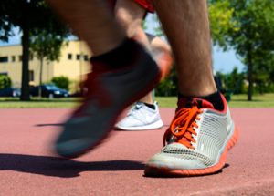 Ankle injuries are often thought of as sports injuries. But you don’t have to be an athlete or even a “weekend warrior” to turn your ankle and hurt it. Something as simple as walking on an uneven surface can cause a painful, debilitating sprain.
+ See More +
Ankle injuries can happen to anyone at any age. However, men between 15 and 24 years old have higher rates of ankle sprain, compared to women older than age 30 who have higher rates than men. Half of all ankle sprains occur during an athletic activity. Every day in the U.S., 25,000 people sprain their ankle. And more than 1 million people visit emergency rooms each year because of ankle injuries. The most common ankle injuries are sprains and fractures, which involve ligaments and bones in the ankle. But you can also tear or strain a tendon. What Kinds of Ankle Injuries Are There? Sprains, Strains, and Fractures Ankle injuries are defined by the kind of tissue — bone, ligament, or tendon — that’s damaged. The ankle is where three bones meet — the tibia and fibula of your lower leg with the talus of your foot. These bones are held together at the ankle joint by ligaments, which are strong elastic bands of connective tissue that keep the bones in place while allowing normal ankle motion. Tendons attach muscles to the bones to do the work of making the ankle and foot move, and help keep the joints stable. A fracture describes a break in one or more of the bones. A sprain is the term that describes damage to ligaments when they are stretched beyond their normal range of motion. A ligament sprain can range from many microscopic tears in the fibers that comprise the ligament to a complete tear or rupture. A strain refers to damage to muscles and tendons as a result of being pulled or stretched too far. Muscle and tendon strains are more common in the legs and lower back. In the ankle, there are two tendons that are often strained. These are the peroneal tendons, and they stabilize and protect the ankle. They can become inflamed as a result of overuse or trauma. Acute tendon tears result from a sudden trauma or force. The inflammation of a tendon is called tendinitis. Microscopic tendon tears that accumulate over time, because of being repeatedly over stretched, and don’t heal properly lead to a condition called tendinosis. Tendons can also rupture. Subluxation refers to a tendon that slips out of place.
article by WebMD
Tarsal Tunnel Syndrome
What is tarsal tunnel syndrome?
Tarsal tunnel syndrome is a condition caused by repeated pressure that results in damage on the posterior tibial nerve. Your tibial nerve branches off of the sciatic nerve and is found near your ankle.
+ See More +
The tibial nerve runs through the tarsal tunnel, which is a narrow passageway inside your ankle that is bound by bone and soft tissue. Damage of the tibial nerve typically occurs when the nerve is compressed as a result of consistent pressure.What are the symptoms of tarsal tunnel syndrome?
Symptoms
People with tarsal tunnel syndrome may experience pain, numbness, or tingling. This pain can be felt anywhere along the tibial nerve, but it’s also common to feel pain in the sole of the foot or inside the ankle. This can feel like: sharp, shooting pains pins and needles an electric shock a burning sensation Symptoms vary greatly depending on each individual. Some people experience symptoms that progress gradually, and some experience symptoms that begin very suddenly. Pain and other symptoms are often aggravated by physical activity. But if the condition is long-standing, some people even experience pain or tingling at night or when resting.
Causes
Tarsal tunnel syndrome results from compression of the tibial nerve, and it’s often caused by other conditions. Causes can include: severely flat feet, because flattened feet can stretch the tibial nerve benign bony growths in the tarsal tunnel varicose veins in the membrane surrounding the tibial nerve, which cause compression on the nerve inflammation from arthritis lesions and masses like tumors or lipomas near the tibial nerve injuries or trauma, like an ankle sprain or fracture — inflammation and swelling from which lead to tarsal tunnel syndrome diabetes, which makes the nerve more vulnerable to compression.
article by health line
READY FOR AN APPOINTMENT?
HOURS
Monday: 7:00AM - 6:00PM
Tuesday: 12:00AM - 5:00PM
Wednesday: 7:00 AM - 6:00PM
Thursday: 8:00AM - 5:00PM
Friday: 7:00AM - 6:00PM
Saturday: 7:00AM - 11:00AM
Sunday: CLOSED
HOURS
Monday: 7:00AM - 6:00PM
Tuesday: 12:00AM - 5:00PM
Wednesday: 7:00 AM - 6:00PM
Thursday: 8:00AM - 5:00PM
Friday: 7:00AM - 6:00PM
Saturday: 7:00AM - 11:00AM
Sunday: CLOSED
1245 Glen Dr Millersburg, OH 44654
Phone: 330-674-0444